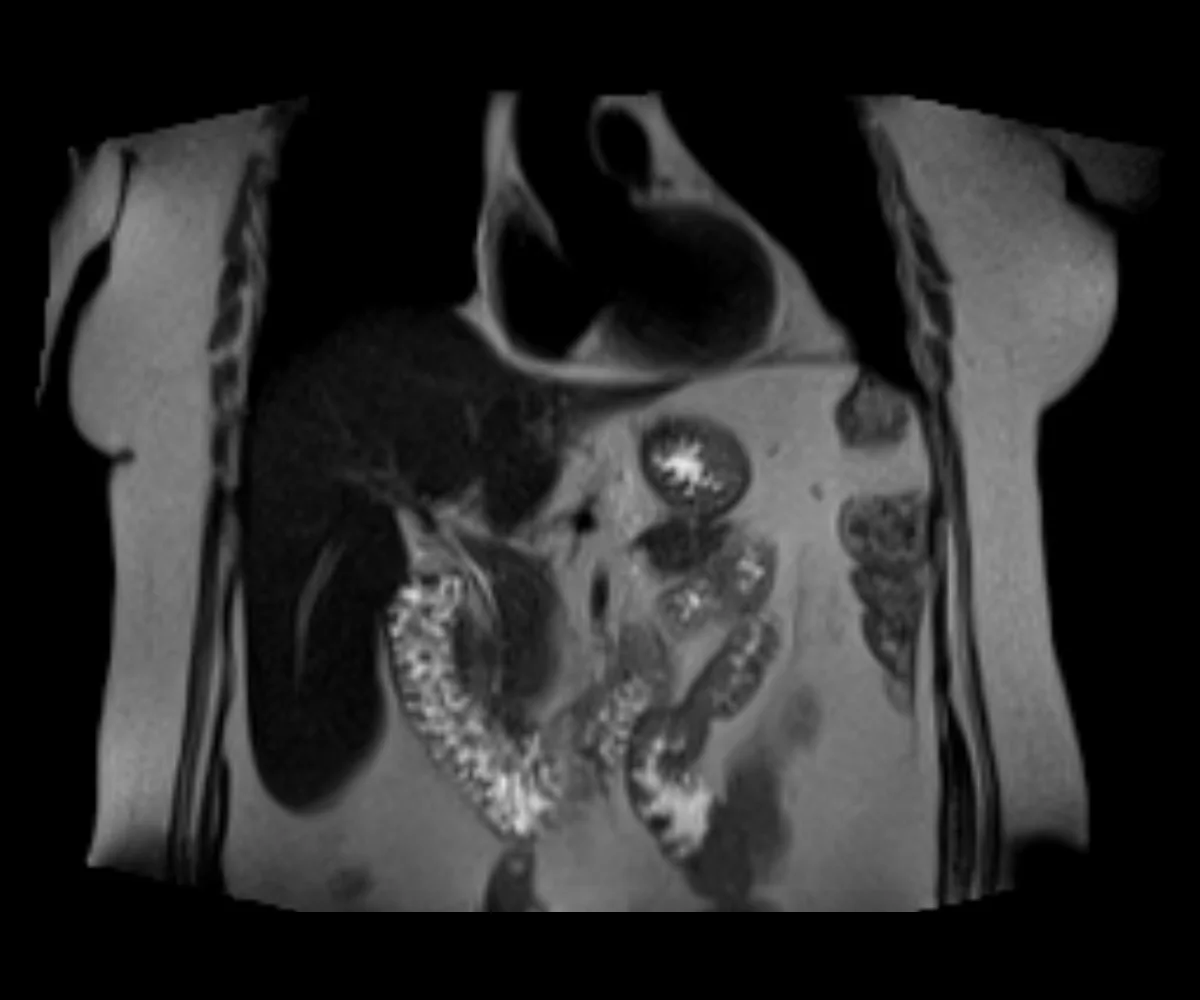
How to Interpret Abdominal MRIs: 3 Essential Techniques
Understanding abdominal MRIs is crucial for diagnosing and treating abdominal conditions effectively. This guide introduces you to the MRI's advantages over X-rays and explains three key techniques for interpreting these detailed images.
Understanding Abdominal MRIs
An abdominal MRI provides a comprehensive view of the abdomen, offering detailed images of organs, tissues, and potential abnormalities. This section outlines what abdominal MRIs are and their advantages for diagnosing various conditions.

What Do Abdominal MRIs Show?
- Organ Detail: Superior imaging of liver, kidneys, pancreas, and other organs.
- Tissue Characterization: Clear visualization of tissues, useful in diagnosing abnormalities.
- Blood Vessels: Detects issues within blood vessels not visible on X-rays.
- Disease Detection: More effective in identifying infections, tumors, and inflammations.
When Will You Get One?
- Unexplained Abdominal Pain: Especially if not adequately explained by an X-ray.
- Suspected Tumors: Such as liver, pancreatic, or kidney tumors.
- Pre-surgical Planning: Provides a detailed anatomy for surgical planning.
- Monitoring Disease Progression: Ideal for assessing changes over time in chronic conditions.
Techniques to Interpret Abdominal MRIs
Interpreting abdominal MRIs involves a deeper understanding of advanced imaging techniques. Here are three techniques for professionals at different levels of expertise.
1. Utilizing X-ray Interpreter
X-ray Interpreter now extends its AI-driven analysis to MRI images. The process ensures precision:
- Registration: Register on X-ray Interpreter to access AI analysis for MRIs.
- Uploading MRIs: Upload your abdominal MRI images.
- Reviewing Interpretation: Receive the AI-generated interpretation and download your report.
- Professional Consultation: Always advisable to consult with medical professionals for comprehensive understanding.
Please check out our get started guide.
2. Using ChatGPT Plus
ChatGPT Plus now supports MRI image analysis with its latest GPT-4V model, offering detailed and interactive insights:
- Subscription: Subscribe to ChatGPT Plus for advanced image analysis features.
- Uploading MRIs: Interface with GPT-4V on OpenAI to upload your MRI images.
- Requesting Analysis: Engage with the model for a thorough analysis.
- Review and Confirmation: Assess and refine the analysis as needed.
- Professional Validation: Validation by medical experts is recommended.
Read more on our post about using ChatGPT Plus for MRI interpretation.
Alternatively, as several other AI models with vision capabilities emerge, you can also try other models, such as Grok by xAI, Claude by Anthropic, Gemini by Google Deepmind.
3. Mastering MRI Interpretation Yourself
For healthcare professionals aiming to enhance their MRI reading skills, self-learning is invaluable:
- Education: Pursue advanced training in MRI interpretation.
- Practice: Regular practice under expert guidance.
- Resources: Utilize advanced imaging books and online courses.
- Feedback: Seek feedback to refine skills.
- Continuous Learning: Engage in ongoing education to stay current with imaging techniques.
Recommended Resources for Self-Learning:
-
MRI of the Abdomen and Pelvis - Radiology Key: This comprehensive resource provides detailed information on the techniques, protocols, and considerations for abdominal and pelvic MRI, making it an invaluable tool for both beginners and advanced learners.
-
Abdominal MRI Interpretation - YouTube: A visual guide that walks through the interpretation of abdominal MRI images. This video is beneficial for visual learners who prefer step-by-step explanations and demonstrations.
-
Abdomen MRI Video - Mediphany: Offers in-depth video content that explains the intricacies of abdominal MRI, including common pathologies and interpretation strategies, suitable for enhancing practical understanding and application.
Comparative Analysis
Choosing the right technique for interpreting abdominal MRIs is vital for accurate diagnosis. This section compares the three methods:
| Criteria | X-ray Interpreter | ChatGPT Plus | Self-Reading |
|---|---|---|---|
| Accuracy | High (AI-based)1 | High (AI-based)1 | Varies (Skill-dependent) |
| Ease of Use | Easy | Moderate | Challenging |
| Cost | Starting from $2.50 per image | $20 per month | Free (excluding educational costs) |
| Time Efficiency | Fast | Moderate to Fast | Slow to Moderate |
| Learning Curve | Low | Low to Moderate | High |
| Additional Resources | Provided | Partially Provided (through OpenAI) | Self-sourced |
Each method has its strengths and weaknesses, with AI options providing rapid and precise interpretations, while self-reading encourages in-depth learning for medical professionals.
Conclusion
Abdominal MRI interpretation is critical for diagnosing complex abdominal conditions. This guide presents three techniques suited to different professional needs and skill levels. AI methods offer quick, precise interpretations, while self-learning is geared towards those seeking in-depth knowledge.
When selecting a technique, consider your level of expertise, the need for timely interpretation, and the resources available. Ethical and legal standards must always be maintained to ensure patient safety and confidentiality.
Related Articles
- How To Interpret Pelvis MRIs
- How To Interpret Abdominal X-Rays
- How To Interpret Abdomen CT Scans
- How To Interpret Abdominal Ultrasounds
Resources and Further Learning
For further exploration and understanding of abdominal MRI interpretation, consider the following resources:
-
Abdominal MRI Scan - Healthline: Provides a comprehensive overview of what an abdominal MRI scan involves, including preparation, procedure, and potential risks. This resource is ideal for patients looking to understand what to expect during an MRI scan.
-
Abdominal and Pelvic MRI - RadiologyInfo.org: Offers detailed information on the use of MRI for imaging the abdomen and pelvis, including indications, benefits, risks, and what patients can expect during the procedure. This resource is useful for both patients and healthcare professionals.
-
MRI of the Abdomen - Sansum Clinic: This resource provides a detailed explanation of abdominal MRI procedures, including the types of conditions it can diagnose, preparation guidelines, and what to expect during and after the scan. It is geared towards both patients and medical professionals seeking a thorough understanding of the procedure.