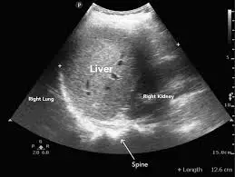
How to interpret abdomen ultrasound: 3 Essential Methods
Abdominal ultrasound is a common imaging technique that uses sound waves to create pictures of the organs and structures inside your abdomen. This guide explains what an abdominal ultrasound can reveal, why it’s used, and how to understand its findings.
What is an Abdominal Ultrasound?
An abdominal ultrasound is a non-invasive imaging test that provides real-time images of the organs and tissues in your abdomen. Unlike X-rays, it uses sound waves and is safe and painless. Let’s explore what this technique involves and what it shows.

What Can Abdominal Ultrasound Show?
- Detailed Organ Imaging: Clear visualization of the liver, gallbladder, kidneys, spleen, pancreas, and major blood vessels.
- Fluid Detection: Can effectively identify fluid collections such as ascites or cysts.
- Abnormality Identification: Helps to detect masses, tumors, gallstones, and kidney stones.
- Real-time Imaging: Allows doctors to observe organ function and blood flow in real-time.
- Guidance for Procedures: Can be used to guide biopsies and other procedures.
Why Might You Need an Abdominal Ultrasound?
- Abdominal Pain: To help identify the cause of unexplained abdominal discomfort.
- Abnormal Blood Tests: When liver or kidney function tests are abnormal.
- Suspected Gallstones: To examine the gallbladder and bile ducts.
- Suspected Enlarged Organs: To examine the liver, spleen or other organs.
- Follow-up Imaging: To monitor the progress of known conditions.
How is an Abdominal Ultrasound Performed?
An abdominal ultrasound is a non-invasive procedure that usually takes about 20-45 minutes. Here's a breakdown of what you can expect:
Preparation
- Clothing: You might be asked to change into a gown. Wear comfortable clothing to make the process easier.
- Fasting: You might be asked to fast (usually for 6-8 hours) before the ultrasound, especially if the gallbladder is being examined. This reduces gas in the intestines for clearer images. Follow the specific instructions given by your doctor or clinic.
- Instructions: Follow any specific instructions from your healthcare provider on how to prepare for the scan.
Procedure
- Gel Application: A clear, water-based gel is applied to your abdomen. This gel ensures good contact between the transducer and your skin.
- Transducer Movement: The sonographer will gently move the transducer over the gelled area of your abdomen. This transducer emits sound waves that create images of your internal organs on the monitor.
- Image Acquisition: The sonographer will view the images in real-time and capture images. You might be asked to hold your breath or change positions to help visualize the organs.
- Patient Comfort: The sonographer will do their best to make you comfortable during the procedure. Tell them if you feel any discomfort.

After the Ultrasound
- Gel Removal: The gel is wiped off, and you can get dressed.
- Results: The ultrasound images will be reviewed by a radiologist. Your physician will then discuss the results with you at a follow-up appointment.
The procedure is painless and does not involve any radiation. If you have any questions or concerns, don’t hesitate to ask the sonographer or your healthcare provider.
How to Interpret Abdominal Ultrasound Results
Understanding your ultrasound results is key to managing your health effectively. Here's an overview of some common ways you can get help interpreting the results.
1. Utilizing X-ray Interpreter
X-ray Interpreter now analyzes ultrasound images with its AI-driven analysis. Here’s how to use it:
- Registration: Sign up on X-ray Interpreter to use our AI for ultrasound analysis.
- Uploading Ultrasounds: Upload your abdominal ultrasound images.
- Reviewing Interpretation: Receive the AI-generated interpretation, including a report.
- Consultation: Always consult with your physician for comprehensive diagnosis.
Check our get started guide for more details.
2. Using ChatGPT Plus
ChatGPT Plus, with its advanced GPT-4V model, can assist in analyzing ultrasound images:
- Subscription: Subscribe to ChatGPT Plus for advanced analysis.
- Uploading Ultrasounds: Upload your ultrasound images on the OpenAI platform.
- Request Analysis: Ask the AI to interpret your ultrasound images and give you a report.
- Review and Validate: Review the results and confirm its accuracy with a healthcare professional.
Find out more in our blog on using ChatGPT Plus for medical image interpretation.
Alternatively, as several other AI models with vision capabilities emerge, you can also try other models, such as Grok by xAI, Claude by Anthropic, Gemini by Google Deepmind.
3. Understanding the Basics Yourself
While not a replacement for medical professionals, understanding some basics can help you better comprehend the results and prepare questions for your doctor.
- Learn Anatomy: Familiarize yourself with the basic anatomy of the abdominal organs.
- Read Simple Guides: Many online sources can help you understand common findings in ultrasounds.
- Ask Questions: Note any unfamiliar terms and ask your healthcare provider during the follow up.
- Seek Expert Guidance: Always validate your understanding with a medical professional.
Comparing the Different Approaches
Let's compare the different methods for interpreting abdominal ultrasounds:
| Criteria | X-ray Interpreter | ChatGPT Plus | Self-Reading |
|---|---|---|---|
| Accuracy | High (AI-based)1 | High (AI-based)1 | Varies (Skill-dependent) |
| Ease of Use | Easy | Moderate | Challenging |
| Cost | Starting from $2.50 per image | $20 per month | Free (excluding educational costs) |
| Time Efficiency | Fast | Moderate to Fast | Slow to Moderate |
| Learning Curve | Low | Low to Moderate | High |
| Additional Resources | Provided | Partially Provided (through OpenAI) | Self-sourced |
Each method has its advantages and disadvantages. AI-driven options are fast and precise, while basic understanding aids in better patient-doctor communication.
Conclusion
Abdominal ultrasounds are a valuable tool for diagnosing various medical conditions. This guide has introduced you to the use of ultrasound technology, the images they produce, and ways you can better understand your results through AI tools and self-guided research.
When choosing a method, consider your specific needs, desired level of understanding, and the resources available. Always adhere to privacy standards and seek expert medical validation.
Related Articles
- How To Interpret Pelvic Ultrasounds
- How To Interpret Abdominal X-Rays
- How To Interpret Abdomen CT Scans
- How To Interpret Abdominal MRIs