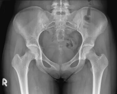
How to interpret pelvic X-rays: 3 Essential Methods
Introduction
Interpreting pelvic X-rays is a crucial skill in many medical and healthcare fields as it provides vital information for diagnosing and treating patients. This guide aims to introduce readers to pelvic X-rays and provide three essential methods to interpret them.
Understanding Pelvic X-rays (PXRs)
A pelvic X-ray is a common imaging test that captures images of the pelvic bones, including the hip bones, sacrum, and coccyx. This section will help you understand what pelvic X-rays are and when you might need one.

What Do Pelvic X-rays Show?
- Bone Structures: Condition of the pelvic bone structure including the hip bones, sacrum, and coccyx.
- Joint Spaces: The sacroiliac joints and the symphysis pubis joint space.
- Foreign Objects: Locating foreign objects like swallowed items or metal fragments.
- Fractures and Dislocations: Identifying fractures and dislocations in the pelvic region.
When Will You Get One?
- Symptom Investigation: Persistent pelvic or lower back pain, hip pain, or after a pelvic injury.
- Routine Check-ups: Especially for individuals with chronic pelvic or hip conditions.
- Pre-surgical Assessment: Before certain surgeries to check the general health of your pelvic bones and joints.
- Occupational Health Screenings: Monitoring bone health in individuals exposed to harmful substances.
Methods to Interpret Pelvic X-rays
Interpreting pelvic X-rays is a nuanced process that can be approached in various ways depending on your expertise, needs, and resources. Below, we delve into three distinct methods that cater to different skill levels and circumstances.
1. Utilizing X-ray Interpreter
X-ray Interpreter is a robust online platform designed to provide quick and accurate interpretations of pelvic X-rays through AI technology. The steps to obtain an AI-generated analysis of your X-ray images remain the same:
- Registration: Sign up on X-ray Interpreter to access the AI-based analysis.
- Uploading X-rays: Upload your pelvic X-ray images onto the platform.
- Reviewing Interpretation: Review the AI-generated interpretation and download the report.
- Seeking Professional Advice: If necessary, consult with medical professionals to understand the interpretation in a clinical context.
Please check out our get started guide.
2. Using ChatGPT Plus
ChatGPT Plus also offers insightful analysis of pelvic X-ray images, providing an interactive experience for users. The steps to obtain a detailed analysis of your X-ray images using ChatGPT Plus are as follows:
- Subscription: Subscribe to ChatGPT Plus to access GPT-4V for image analysis.
- Uploading X-rays: Navigate to the GPT-4V interface on the OpenAI platform and upload your pelvic X-ray images.
- Requesting Analysis: Request an analysis of the X-ray images by inputting natural language commands or questions.
- Reviewing and Confirming Analysis: Review the analysis provided, iterate to get more detailed or specific information if necessary.
- Consulting Professionals: Seek advice from medical professionals to validate the analysis provided by GPT-4V.
Please read our post on how to use ChatGPT Plus for X-ray analysis to learn more.
Alternatively, as several other AI models with vision capabilities emerge, you can also try other models, such as Grok by xAI, Claude by Anthropic, Gemini by Google Deepmind.
3. Reading X-rays by Yourself
Self-reading is a traditional method that relies on individual expertise and resources, ideal for medical professionals looking to hone their interpretative skills in pelvic X-rays:
- Education: Acquire basic knowledge and training on reading and interpreting pelvic X-rays from reputable sources or through coursework.
- Practice: Practice interpreting pelvic X-rays under the guidance of experienced professionals.
- Resources: Utilize books, online resources, and medical literature to enhance your understanding and skills in interpreting pelvic X-rays.
- Seeking Feedback: Obtain feedback from knowledgeable professionals to improve your interpretation skills.
- Continuous Learning: Continuously update your knowledge and skills by reading recent medical literature, attending workshops, and engaging in discussions with professionals.
Recommended Resources for Self-Reading:
- Radiopaedia - Pelvic Radiograph: Offers a systematic review for identifying fractures and examining joint spaces in pelvic X-rays.
- WikEM - Pelvic X-ray Interpretation: Provides a checklist for evaluating symmetry, joint spaces, and integrity of cortical lines in pelvic X-rays.
- GrepMed - Pelvic X-Ray Anatomy and Interpretation Checklist: A detailed checklist covering various aspects of pelvic X-ray anatomy and interpretation.
Comparative Analysis
Depending on your circumstances and level of expertise, you may find one method more suitable than the others.
The table below provides a quick overview of how each method stacks up against the others across various criteria:
| Criteria | X-ray Interpreter | ChatGPT Plus | Self-Reading |
|---|---|---|---|
| Accuracy | Mostly High (AI-based)1 | Mostly High (AI-based)1 | Varies (Skill-dependent) |
| Ease of Use | Easy | Moderate | Challenging |
| Cost | Starting from $2.50 per image | $20 per month | Free (excluding educational costs) |
| Time Efficiency | Fast | Moderate to Fast | Slow to Moderate |
| Learning Curve | Low | Low to Moderate | High |
| Additional Resources | Provided | Partially Provided (through OpenAI) | Self-sourced |
Conclusion
This guide illuminated three divergent methods for interpreting pelvic X-rays: leveraging X-ray Interpreter, employing ChatGPT Plus, and self-reading. Each technique caters to distinct levels of expertise and circumstances, offering a spectrum of options for individuals and professionals alike.
X-ray Interpreter and ChatGPT Plus harness the prowess of artificial intelligence to furnish rapid and precise interpretations, making them suitable selections for those requiring immediate insights, irrespective of their medical background. Conversely, self-reading is a more conventional approach, ideal for medical professionals desiring to refine their interpretative acumen.
The comparative analysis furnished herein aims to aid in making an enlightened decision based on individual needs, technical proficiency, and available resources. While AI-based methods proffer a high level of accuracy and user-friendliness, self-reading provides an avenue for continuous learning and professional advancement.
Moreover, the guide underscored several resources to facilitate self-education for those engrossed in the self-reading method. These resources offer a treasure trove of information and practice materials to augment one's skills in pelvic X-ray interpretation.
In a swiftly evolving medical landscape, staying abreast with the latest technologies and methodologies is indispensable for delivering accurate and timely patient care. Regardless of the method chosen, adherence to legal guidelines and ethical practices is paramount to ensure the privacy, safety, and wellbeing of individuals.
This guide serves as a springboard in exploring the various methods of pelvic X-ray interpretation, with the aspiration of aiding individuals and professionals in navigating this crucial aspect of medical diagnostics. The choice between these methods ultimately hinges on personal preferences, professional aspirations, and the specific circumstances at hand.
Related Articles
- How To Interpret Abdominal X-Rays
- How To Interpret Pelvis CT Scans
- How To Interpret Pelvic MRIs
- How To Interpret Pelvic Ultrasounds
Resources and Further Learning
For those keen on delving deeper into pelvic X-ray interpretation, a plethora of resources are available online and in print. Whether you are a seasoned professional aiming to refresh your knowledge or a student eager to learn, these resources offer invaluable insights and structured approaches to interpreting pelvic X-rays:
-
Pelvic Radiograph - An Approach on Radiopaedia:
- This resource provides a structured approach to interpreting pelvic radiographs, with a focus on identifying fractures and assessing joint spaces.
-
Pelvic X-Ray Interpretation on WikEM:
- WikEM provides a checklist and detailed guide on measurements and views essential for accurate pelvic X-ray interpretation, aiding in identifying asymmetry and other abnormalities.
-
How to Read Pelvic X-Rays on International Emergency Medicine Education:
- This guide introduces the Young-Burgess system for identifying pelvic ring disruptions and provides insight into interpreting various types of injuries visible on pelvic X-rays.
The aforementioned resources provide a wide array of learning materials, tutorials, and checklists, designed to enhance your skills and understanding in pelvic X-ray interpretation. Each resource caters to different levels of expertise, aiding in the continuous learning and professional development of individuals keen on mastering the art of pelvic X-ray interpretation.