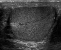
How to interpret scrotal ultrasounds: 3 Essential Methods
A scrotal ultrasound is a key imaging technique using sound waves to examine the testicles and surrounding tissues. This guide explains what this ultrasound can reveal, why it's necessary, and how to interpret its findings.
What is a Scrotal Ultrasound?
A scrotal ultrasound is a non-invasive imaging test that provides real-time images of the testicles, epididymis, and surrounding structures. Using sound waves, it’s a safe, painless way to assess the male reproductive organs. Let's explore what it shows and the process.

What Can a Scrotal Ultrasound Show?
- Detailed Testicular Imaging: Clear visualization of the testicles, epididymis, and spermatic cords.
- Fluid Detection: Detects fluid collections like hydroceles or varicoceles.
- Abnormality Identification: Helps identify masses, tumors, cysts, and signs of inflammation or infection.
- Blood Flow Assessment: Evaluates blood flow to the testicles, crucial for diagnosing torsion.
- Real-time Imaging: Allows doctors to observe the structures in real-time.
Why Might You Need a Scrotal Ultrasound?
- Testicular Pain or Swelling: To identify the cause of discomfort or enlargement.
- Palpable Mass or Lump: To assess any abnormalities felt during self-exams or by a doctor.
- Infertility Concerns: To evaluate the testicular structure and identify potential issues.
- Trauma to the Scrotum: To assess damage after injury.
- Suspected Torsion: To rule out or confirm testicular torsion, a medical emergency.
How is a Scrotal Ultrasound Performed?
A scrotal ultrasound is a quick, painless, and non-invasive procedure. Here’s what you can typically expect:
-
Preparation: You will be asked to remove any clothing from the waist down and will be provided with a gown or drape. You’ll then lie on your back on an examination table.
-
Gel Application: A clear, water-based gel will be applied to the skin of your scrotum. This gel helps the transducer (the handheld device that emits sound waves) make good contact with your skin and allows for better transmission of sound waves.
-
Transducer Movement: The sonographer (the trained professional performing the ultrasound) will then gently move the transducer over the gel-covered area of your scrotum. They will apply slight pressure as needed to get the best images of the testicles, epididymis, and surrounding structures.
-
Image Acquisition: As the transducer is moved, real-time images are displayed on a monitor. The sonographer may take still images or video clips of particular areas. You might be asked to hold still or change your position slightly to get the best views.
-
Procedure Duration: The entire procedure usually takes about 15-30 minutes.
-
Post Procedure: After the procedure, the gel will be wiped off, and you can get dressed and go about your day. The images are then usually reviewed by a radiologist, who will provide a report to your doctor.

How to Interpret Scrotal Ultrasound Results
Understanding your ultrasound results is crucial for managing your health. Here's how you can get help interpreting them.
1. Utilizing X-ray Interpreter
X-ray Interpreter now provides AI-driven analysis for ultrasound images, including scrotal ultrasounds. Here’s how to use it:
- Registration: Sign up on X-ray Interpreter to use AI for ultrasound analysis.
- Uploading Ultrasounds: Upload your scrotal ultrasound images.
- Reviewing Interpretation: Get AI-generated analysis and a detailed report.
- Consultation: Always consult with your doctor for comprehensive diagnosis.
Check our get started guide for more details.
2. Using ChatGPT Plus
ChatGPT Plus, using GPT-4V, can help analyze ultrasound images:
- Subscription: Subscribe to ChatGPT Plus for advanced analysis.
- Uploading Ultrasounds: Upload your images on the OpenAI platform.
- Request Analysis: Ask the AI to interpret your ultrasound and provide a report.
- Review and Validate: Review the results with a healthcare professional.
Find out more in our blog on using ChatGPT Plus for medical image interpretation.
Alternatively, as several other AI models with vision capabilities emerge, you can also try other models, such as Grok by xAI, Claude by Anthropic, Gemini by Google Deepmind.
3. Understanding the Basics Yourself
While not a substitute for medical advice, understanding basic concepts can help you better comprehend results.
- Learn Anatomy: Familiarize yourself with the anatomy of the testicles, epididymis, and surrounding structures.
- Read Guides: Many online resources explain common ultrasound findings.
- Ask Questions: Note unfamiliar terms and clarify with your healthcare provider.
- Seek Expert Guidance: Always validate your understanding with a medical professional.
Comparing the Different Approaches
Let's compare methods for interpreting scrotal ultrasounds:
| Criteria | X-ray Interpreter | ChatGPT Plus | Self-Reading |
|---|---|---|---|
| Accuracy | High (AI-based)1 | High (AI-based)1 | Varies (Skill-dependent) |
| Ease of Use | Easy | Moderate | Challenging |
| Cost | Starting from $2.50 per image | $20 per month | Free (excluding educational costs) |
| Time Efficiency | Fast | Moderate to Fast | Slow to Moderate |
| Learning Curve | Low | Low to Moderate | High |
| Additional Resources | Provided | Partially Provided (through OpenAI) | Self-sourced |
AI-driven methods offer speed and precision, while basic knowledge promotes better communication with doctors.
Conclusion
Scrotal ultrasounds are essential for diagnosing male reproductive issues. This guide has explained their use, the images they produce, and methods for understanding results using AI tools and self-education.
When choosing a method, consider your specific needs and resources. Always prioritize privacy and seek professional validation.