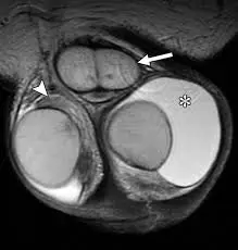
How to Interpret Scrotal MRIs: 3 Essential Techniques
Scrotal MRI is a powerful imaging technique used to examine the testicles and surrounding tissues. This guide explains what a scrotal MRI can reveal, why it's used, and how to interpret its findings.
What is a Scrotal MRI?
A scrotal MRI (Magnetic Resonance Imaging) is a non-invasive imaging test that uses magnetic fields and radio waves to produce detailed images of the scrotum. It provides superior soft tissue contrast compared to ultrasound and is particularly useful for evaluating abnormalities.

What Can Scrotal MRI Show?
- Detailed Tissue Imaging: Clear visualization of the testicles, epididymis, spermatic cord, and surrounding tissues.
- Tumor Detection: Effective in identifying testicular tumors and other masses.
- Inflammation Assessment: Helps in evaluating epididymitis, orchitis, and other inflammatory conditions.
- Vascular Abnormalities: Can detect varicoceles, testicular torsion, and other vascular issues.
- Cryptorchidism Evaluation: Useful in locating undescended testicles.
- Post-Trauma Assessment: Helps in evaluating injuries to the scrotum.
Why Might You Need a Scrotal MRI?
- Testicular Pain or Swelling: To investigate the cause of unexplained pain or swelling in the scrotum.
- Palpable Mass: When a mass is felt during a physical exam.
- Abnormal Ultrasound: To further evaluate abnormalities found during scrotal ultrasound.
- Suspected Testicular Cancer: To help stage testicular tumors.
- Infertility Evaluation: As part of an evaluation for male infertility.
- Trauma Evaluation: To assess the extent of injuries after trauma.
How to Interpret Scrotal MRI Results
Interpreting your MRI results is crucial for understanding your diagnosis. Here are methods to help you understand the findings.
1. Utilizing X-ray Interpreter
X-ray Interpreter offers AI-driven analysis for scrotal MRI interpretation. Here’s how to use it:
- Sign Up: Register at X-ray Interpreter to use our AI.
- Upload Images: Upload your scrotal MRI images.
- Get Interpretation: Receive an AI-generated report.
- Consult Your Physician: Always seek a professional diagnosis.
See our guide for more details.
2. Using ChatGPT Plus
ChatGPT Plus, with its advanced GPT-4V model, can analyze MRI images:
- Subscribe: Subscribe to ChatGPT Plus.
- Upload Images: Upload your MRI images to the OpenAI platform.
- Request Analysis: Ask the AI to interpret your MRI.
- Review and Validate: Consult a healthcare provider for accuracy.
Find out more in our blog on using ChatGPT Plus for medical image analysis.
Alternatively, as several other AI models with vision capabilities emerge, you can also try other models, such as Grok by xAI, Claude by Anthropic, Gemini by Google Deepmind.
3. Understanding the Basics Yourself
Understanding the basics of MRI can aid in understanding your report.
- Learn Scrotal Anatomy: Familiarize yourself with the basics.
- Read Simple Guides: There are many resources available online.
- Ask Questions: Make note of medical terms and consult with your doctor.
- Seek Expert Guidance: Validate your understanding with a medical professional.
Comparing Different Approaches
| Criteria | X-ray Interpreter | ChatGPT Plus | Self-Reading |
|---|---|---|---|
| Accuracy | Mostly High (AI-based)1 | Mostly High (AI-based)1 | Varies (Skill-dependent) |
| Ease of Use | Easy | Moderate | Challenging |
| Cost | Starting from $2.50 per image | $20 per month | Free (excluding educational costs) |
| Time Efficiency | Fast | Moderate to Fast | Slow to Moderate |
| Learning Curve | Low | Low to Moderate | High |
| Additional Resources | Provided | Partially Provided (through OpenAI) | Self-sourced |
Each method offers benefits. AI provides speed and accuracy, while basic knowledge assists in patient-doctor discussions.
Conclusion
Scrotal MRI is a crucial diagnostic tool for many conditions. This guide introduced you to the benefits, interpretation, and use of AI and self-guided learning. Always consult with a doctor for a comprehensive diagnosis.