How to interpret leg X-rays: 3 Essential Methods
Introduction
Mastering leg X-ray analysis is a vital skill in orthopedics and sports medicine. It plays a crucial role in diagnosing fractures, joint disorders, and other lower extremity conditions. This guide aims to familiarize practitioners and students with the fundamentals of interpreting leg radiographs. Focusing on both long bone and joint imaging, we delve into the intricacies of leg radiography interpretation. Suitable for both experienced medical professionals and beginners, our guide simplifies complex concepts, ensuring a solid understanding of this essential diagnostic tool.
Exploring Leg X-rays: Comprehensive Guide on Long Bone and Joint Imaging
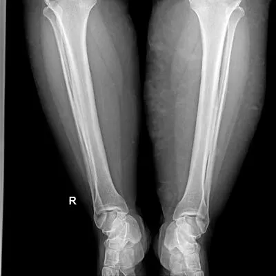
Leg X-rays, including long bone and joint imaging, are vital radiographic tools in orthopedic care. These imaging techniques provide detailed views of the femur, tibia, fibula, knee, ankle, and foot. This section explains what leg X-rays are and their critical role in orthopedic diagnosis. Understanding the specific features of long bone and joint X-rays, including their applications and advantages, allows healthcare professionals to make informed decisions in various clinical situations, from fracture assessment to joint disease evaluation.
Long Bone vs. Joint X-rays: Understanding Different Types
Leg X-rays can be categorized into long bone and joint imaging. Each type has a distinct purpose in assessing lower extremity health.
Long Bone X-rays: These images focus on the femur, tibia, and fibula, providing details on bone integrity, alignment, and the presence of fractures or deformities. They are crucial for evaluating traumatic injuries, monitoring bone healing, and planning orthopedic surgeries.
Joint X-rays: These offer a detailed view of the knee, ankle, and foot joints, essential for diagnosing arthritis, ligament injuries, and other joint disorders. Weight-bearing X-rays of the knee or ankle, for instance, are particularly valuable for assessing joint alignment and degenerative changes.
Indications for Leg X-rays: When and Why They Are Needed
Leg X-rays are critical diagnostic tools for specific lower extremity conditions and scenarios. Common reasons for requiring a leg X-ray include:
- Symptom Investigation: For persistent leg pain, swelling, or mobility issues, leg X-rays provide a clear view of underlying skeletal problems.
- Trauma Assessment: Essential for evaluating injuries from accidents or sports.
- Pre-Surgical Planning: Crucial before orthopedic procedures to thoroughly assess bone and joint health.
- Chronic Condition Monitoring: Invaluable for managing long-term conditions like osteoarthritis or osteoporosis.
Recognizing when and why leg X-rays are necessary is key for healthcare professionals and patients in managing lower extremity health effectively.
Expert Techniques for Interpreting Leg X-rays
Interpreting leg X-rays involves a nuanced process, varying based on expertise and available tools. We present three effective methods:
1. AI-Assisted Analysis with X-ray Interpreter
X-ray Interpreter utilizes cutting-edge AI technology for fast, accurate interpretations of leg X-ray images. It's user-friendly and ideal for efficient, reliable analysis.
Following these steps, you can easily obtain an AI-generated analysis of your leg X-ray images:
- Registration: Register on X-ray Interpreter to access the AI-powered analysis.
- Uploading X-rays: Upload your leg X-ray images onto the platform.
- Reviewing Interpretation: Examine the AI-generated interpretation and download the report.
- Consulting Medical Professionals: If necessary, discuss the interpretation with healthcare professionals to understand its clinical significance.
Please check out our get started guide.
2. Interactive Analysis with ChatGPT Plus
ChatGPT Plus employs the advanced GPT-4V model to provide insightful analysis of leg X-ray images. This method offers an interactive experience, allowing for communication with the AI to tailor the analysis to specific needs:
- Subscription: Subscribe to ChatGPT Plus for access to the GPT-4V image analysis feature.
- Uploading X-rays: Go to the GPT-4V interface on the OpenAI platform and upload your leg X-ray images.
- Requesting Analysis: Input natural language commands or questions to request an analysis of the X-ray images.
- Reviewing and Confirming Analysis: Evaluate the analysis provided, and refine it for more detailed or specific information if needed.
- Consulting Medical Experts: Seek advice from healthcare professionals to validate the analysis from GPT-4V.
Please read our post on how to use ChatGPT Plus for leg X-ray analysis to learn more.
Alternatively, as several other AI models with vision capabilities emerge, you can also try other models, such as Grok by xAI, Claude by Anthropic, Gemini by Google Deepmind.
3. Self-Reading Leg X-rays
Self-reading is a traditional method relying on individual expertise. It's ideal for medical professionals seeking to enhance their interpretative skills. This method requires solid knowledge of orthopedic radiology and a commitment to continuous learning:
- Education: Gain basic knowledge and training in reading and interpreting leg X-rays from reputable sources or coursework.
- Practice: Practice interpreting leg X-rays under experienced professionals' guidance.
- Resources: Use books, online resources, and medical literature to deepen understanding and skills in interpreting leg X-rays.
- Feedback: Get feedback from knowledgeable professionals to improve interpretation skills.
- Continuous Learning: Regularly update knowledge and skills by engaging in recent medical literature, workshops, and professional discussions.
Recommended Resources for Self-Reading:
-
X-Ray Exam: Lower Leg (Tibia and Fibula) - Nemours KidsHealth:
- This resource provides comprehensive information about lower leg X-rays, particularly focusing on the tibia and fibula. It explains the procedure, the purpose of the X-ray, and what can be learned from the images. Ideal for understanding the basics of lower leg radiography and its clinical significance.
-
Lower Extremity X-Ray | Cedars-Sinai:
- Cedars-Sinai offers a detailed overview of lower extremity X-rays, covering the procedure and its uses in medical diagnosis and treatment.
-
Trauma X-ray - Lower limb - Radiology Masterclass:
- This tutorial from Radiology Masterclass focuses on the X-ray appearances of lower limb trauma. It includes examples of common injuries and normal images for comparison, making it suitable for clinicians in emergency departments requiring knowledge of trauma X-ray interpretation.
Comparative Review of Leg X-ray Interpretation Methods
Selecting the appropriate method for interpreting leg X-rays is crucial for accurate diagnosis and effective patient care. In this comparative analysis, we assess the three methods - AI-assisted interpretation with X-ray Interpreter, interactive analysis with ChatGPT Plus, and traditional self-reading. Our comparison focuses on accuracy, ease of use, cost, time efficiency, and availability of additional resources. This evaluation aims to provide a comprehensive understanding of each method's strengths and limitations, assisting you in choosing a method suited to your specific needs and expertise in leg X-ray interpretation.
| Criteria | X-ray Interpreter | ChatGPT Plus | Self-Reading |
|---|---|---|---|
| Accuracy | Mostly High (AI-based)1 | Mostly High (AI-based)1 | Varies (Skill-dependent) |
| Ease of Use | Easy | Moderate | Challenging |
| Cost | Starting from $2.50 per image | $20 per month | Free (excluding educational costs) |
| Time Efficiency | Fast | Moderate to Fast | Slow to Moderate |
| Learning Curve | Low | Low to Moderate | High |
| Additional Resources | Provided | Partially Provided (through OpenAI) | Self-sourced |
Summing Up: The Importance of Accurate Leg X-ray Analysis
Interpreting leg X-rays is a key skill in orthopedics, playing a significant role in diagnosing and managing various lower extremity conditions. This guide has introduced three different methods for leg X-ray interpretation: utilizing X-ray Interpreter, using ChatGPT Plus, and self-reading. Each approach caters to varying levels of expertise and circumstances, offering diverse options for individuals and professionals.
As the field of orthopedics continues to evolve, keeping abreast of the latest technologies and methods is vital for providing precise and timely patient care. Regardless of the chosen method, adherence to legal and ethical guidelines is essential to ensure the privacy, safety, and wellbeing of patients.
This guide serves as a foundation for exploring the diverse methods of leg X-ray interpretation, aiming to aid individuals and professionals in this critical aspect of orthopedic diagnostics. The choice among these methods will ultimately depend on personal preferences, professional goals, and specific situations.
Related Articles
Resources and Further Learning
For those seeking deeper insights into leg X-ray interpretation, various online resources are available. These resources offer valuable knowledge and structured approaches to interpreting leg X-rays, enhancing your skills and understanding in this important aspect of orthopedic diagnostics.
-
X-rays of the Extremities | Johns Hopkins Medicine
- This resource from Johns Hopkins Medicine provides a comprehensive overview of X-rays for the extremities, discussing their uses in diagnosing various conditions including tumors, infections, and bone injuries.
-
- UCSF Health offers detailed information on extremity X-rays, covering how the test is performed, its purposes, and what the results might indicate. This is a valuable resource for understanding the practical aspects and clinical relevance of extremity X-rays.