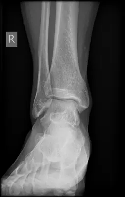
How to interpret ankle X-rays: 3 Essential Methods
Introduction
The interpretation of ankle X-rays is a critical skill in orthopedics and various medical fields, providing essential information for diagnosing and managing patient conditions. This guide will introduce you to ankle X-rays and explain key methods for their interpretation.
Ankle injuries are common and can occur in various settings, such as sports, work, or daily activities. These range from minor sprains and strains to more serious fractures, dislocations, or ligament tears. Ankle X-rays are vital in assessing these injuries, determining the alignment of bones, and planning surgical or conservative treatments. They also play a significant role in monitoring healing progress and ensuring effective recovery.
Understanding Ankle X-rays
Ankle X-rays are common radiographic exams that reveal details of the bones, joints, and soft tissues of the ankle. This section helps you understand ankle X-rays and their indications.

What Do Ankle X-rays Show?
- Bone Conditions: Detects or monitors issues like fractures, arthritis, or bone tumors.
- Joint Conditions: Evaluates joint status, including dislocations and arthritis.
- Foreign Objects: Identifies foreign objects such as glass or metal fragments.
- Soft Tissue Conditions: Shows swelling or abnormalities in adjacent soft tissues.
When Are Ankle X-rays Needed?
- Symptom Investigation: Persistent pain, swelling, deformity, or functional impairment.
- Post-Injury Assessment: Following acute injuries to assess the extent of damage.
- Pre-Surgical Planning: Before surgeries to understand the anatomy and pathology.
- Routine Follow-Up: Especially in patients with ongoing joint or bone conditions.
Methods to Interpret Ankle X-rays
Interpreting ankle X-rays requires skill and knowledge, with various approaches available based on expertise and resources. Here, we explore three methods for different levels of proficiency and needs.
1. Utilizing X-ray Interpreter
AI interpretation tools like X-ray Interpreter offer quick and accurate readings of ankle X-rays through advanced algorithms. To use these tools:
- Sign Up: Register on X-ray Interpreter to access AI-based analysis.
- Upload X-rays: Upload your ankle X-ray images to the platform.
- AI Analysis: Receive an AI-generated interpretation and download the report.
- Professional Consultation: Consult medical professionals for clinical understanding of the AI interpretation.
Check our get started guide for more.
2. ChatGPT Plus for Ankle X-ray Analysis
ChatGPT Plus, using the GPT-4V model, offers detailed analysis of ankle X-ray images. It provides an interactive environment for tailored analysis:
- Subscription: Get ChatGPT Plus for GPT-4V image analysis.
- Upload X-rays: Go to the GPT-4V interface on OpenAI and upload your ankle X-ray images.
- Analysis Request: Input commands or questions for AI analysis.
- Review and Refine: Check the AI analysis, refine as needed.
- Professional Verification: Validate the analysis with medical professionals.
Learn more on using ChatGPT Plus for X-ray analysis.
Alternatively, as several other AI models with vision capabilities emerge, you can also try other models, such as Grok by xAI, Claude by Anthropic, Gemini by Google Deepmind.
3. Self-Interpretation of Ankle X-rays
Self-interpretation suits medical professionals enhancing their skills. It requires a solid orthopedic foundation and commitment to ongoing learning:
Essential Resources for Self-Interpretation:
-
Geeky Medics - Ankle X-ray Interpretation
- Offers a comprehensive guide on interpreting ankle X-rays, covering the anatomy of the ankle joint and key principles of X-ray interpretation.
-
Don't Forget the Bubbles - Ankle X-rays
- Provides insights into ankle X-rays, likely including reading techniques and typical findings.
-
Radiopaedia - Ankle Radiograph (an approach)
- Describes a systematic approach to interpreting ankle radiographs, emphasizing a consistent search strategy to avoid common errors in diagnostic radiology.
-
Radiology Masterclass - Trauma X-ray Lower Limb
- Discusses key points on ankle injuries and the standard views used in ankle X-rays, including insights into understanding the talar dome surface and the involvement of bones and ligaments in ankle injuries.
- Education: Gain basic skills in ankle X-ray reading from trusted sources or courses.
- Practice: Interpret X-rays under expert guidance.
- Resources: Use books, online materials, and journals for skill enhancement.
- Feedback: Seek expert opinions to refine interpretations.
- Continuous Education: Stay updated with latest research and participate in professional forums.
Comparative Analysis
Choosing the appropriate method for ankle X-ray interpretation is crucial for precise and timely diagnosis, impacting patient care. We compare X-ray Interpreter, ChatGPT Plus, and Self-Reading, evaluating accuracy, ease of use, cost, efficiency, learning curve, and resource availability. This helps you make an informed choice suited to your needs.
| Criteria | X-ray Interpreter | ChatGPT Plus | Self-Reading |
|---|---|---|---|
| Accuracy | Mostly High (AI-based)1 | Mostly High (AI-based)1 | Varies (Skill-dependent) |
| Ease of Use | Simple | Moderate | Challenging |
| Cost | Variable per image | Monthly subscription | Free (Excluding educational costs) |
| Time Efficiency | Quick | Quick to Moderate | Varies |
| Learning Curve | Low | Moderate | High |
| Additional Resources | Included | Partially Available (via OpenAI) | Self-provided |
Conclusion
Mastering ankle X-ray interpretation is vital in medical and orthopedic practice, aiding in the diagnosis and treatment of various conditions. This guide reviewed three methods: AI-based interpretation, using ChatGPT Plus, and self-interpretation. Each caters to different expertise levels and scenarios, offering a spectrum of options for both professionals and laypersons.
AI-based methods, like X-ray Interpreter and ChatGPT Plus, provide fast and accurate interpretations, ideal for immediate insights. Self-interpretation, however, is more traditional, suitable for medical professionals seeking skill development.
Our comparative analysis assists in choosing the most appropriate method, considering personal preferences, professional objectives, and available resources. While AI methods offer accuracy and convenience, self-interpretation encourages continuous learning and skill advancement.
In addition to outlining each method, the guide offers resources for those interested in self-learning. Keeping up with evolving technologies and techniques is essential in modern medical care. Regardless of the chosen method, adhering to ethical standards and legal guidelines is paramount for patient safety and privacy.
This guide serves as a foundational resource in ankle X-ray interpretation, aiming to assist both individuals and professionals in navigating this essential aspect of medical diagnostics. The choice depends on personal and professional circumstances.
Related Articles
Resources and Further Learning
For those interested in further exploring ankle X-ray interpretation, here are several valuable resources:
-
- Details the components of an ankle series, typically including anteroposterior (AP), mortise, and lateral radiographs, commonly used in emergency departments for evaluating the ankle joint.
-
Orthobullets - Adult Ankle Radiographs
- Provides information on adult ankle radiographs, useful for medical professionals and students.
-
ALiEM - EMRad: Radiologic Approach to the Traumatic Ankle
- A resource providing a radiologic perspective on evaluating traumatic ankle injuries.
These resources cater to varied expertise levels, offering different perspectives and techniques in ankle X-ray interpretation, enriching your knowledge and skills in this critical area of medical diagnostics.