How to Interpret Carotid Ultrasounds: 3 Essential Methods
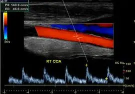
A carotid ultrasound is a non-invasive imaging test that uses sound waves to examine the carotid arteries in your neck, which supply blood to your brain. This guide explains what a carotid ultrasound can reveal, why it's used, and how to interpret its findings.
What is a Carotid Ultrasound?
A carotid ultrasound uses high-frequency sound waves to create images of your carotid arteries. It’s a safe, painless procedure that provides real-time images. Let’s explore what it shows and how it works.

What Can a Carotid Ultrasound Show?
- Blood Flow Analysis: Measures the speed and direction of blood flow in the carotid arteries.
- Plaque Detection: Identifies the buildup of plaque that can cause narrowing or blockages.
- Artery Structure: Visualizes the size and shape of the carotid arteries.
- Stenosis Measurement: Quantifies the degree of narrowing in the arteries (stenosis).
- Real-time Imaging: Provides dynamic views of blood flow patterns.
Why Might You Need a Carotid Ultrasound?
- Stroke or TIA Symptoms: To assess carotid artery health after stroke or transient ischemic attack (TIA).
- Risk Assessment: To identify risk of future stroke in individuals with risk factors.
- Carotid Bruit: To examine if there's an abnormal sound during a physical exam of the neck.
- Follow-up: To monitor known carotid artery conditions.
- Pre-Operative Assessment: To evaluate artery health before certain surgeries.
How is a Carotid Ultrasound Performed?
A carotid ultrasound is a quick, non-invasive procedure. Here's what typically happens:
- Preparation: You'll lie on your back with your neck slightly extended.
- Gel Application: A clear, water-based gel is applied to your neck over the area where the carotid arteries are located. This helps the transducer make better contact with your skin.
- Transducer Placement: A small, handheld device called a transducer is gently moved over your neck. The transducer sends out sound waves.
- Image Capture: The sound waves bounce back, creating images of the arteries on a monitor.
- Doppler Technique: A Doppler mode may be used to analyze blood flow direction and speed.
- Duration: The procedure usually takes about 30 to 60 minutes.

The test is painless, and there are no after-effects. You can resume your normal activities immediately.
How to Interpret Carotid Ultrasound Results
Understanding your carotid ultrasound results is important for managing your health. Here are three ways you can approach interpreting the findings.
1. Utilizing X-ray Interpreter
X-ray Interpreter provides AI-driven analysis for carotid ultrasound images. Here’s how to use it:
- Registration: Sign up on X-ray Interpreter to use our AI for carotid ultrasound analysis.
- Uploading Ultrasounds: Upload your carotid ultrasound images.
- Reviewing Interpretation: Receive the AI-generated interpretation, including a detailed report.
- Consultation: Always consult with your physician for a comprehensive diagnosis.
Check our get started guide for more details.
2. Using ChatGPT Plus
ChatGPT Plus, powered by the advanced GPT-4V model, can assist in analyzing carotid ultrasound images:
- Subscription: Subscribe to ChatGPT Plus for advanced AI analysis.
- Uploading Ultrasounds: Upload your carotid ultrasound images on the OpenAI platform.
- Request Analysis: Ask the AI to interpret your ultrasound images and provide a detailed report.
- Review and Validate: Review the results and confirm their accuracy with a healthcare professional.
Find out more in our blog on using ChatGPT Plus for medical image interpretation.
Alternatively, as several other AI models with vision capabilities emerge, you can also try other models, such as Grok by xAI, Claude by Anthropic, Gemini by Google Deepmind.
3. Understanding the Basics Yourself
While not a substitute for professional medical advice, understanding some basics can help you comprehend the results better and prepare questions for your doctor.
- Learn Carotid Anatomy: Familiarize yourself with the basic anatomy of the carotid arteries.
- Read Simple Guides: Many online resources provide explanations of common findings.
- Ask Questions: Note any unfamiliar terms and ask your healthcare provider for clarification.
- Seek Expert Guidance: Always validate your understanding with a qualified medical professional.
Comparing the Different Approaches
Let's compare the different methods for interpreting carotid ultrasounds:
| Criteria | X-ray Interpreter | ChatGPT Plus | Self-Reading |
|---|---|---|---|
| Accuracy | High (AI-based)1 | High (AI-based)1 | Varies (Skill-dependent) |
| Ease of Use | Easy | Moderate | Challenging |
| Cost | Starting from $2.50 per image | $20 per month | Free (excluding educational costs) |
| Time Efficiency | Fast | Moderate to Fast | Slow to Moderate |
| Learning Curve | Low | Low to Moderate | High |
| Additional Resources | Provided | Partially Provided (through OpenAI) | Self-sourced |
Each method offers its own set of advantages and disadvantages. AI-driven approaches are fast and precise, while a basic understanding can help in better patient-doctor communication.
Conclusion
Carotid ultrasounds are crucial for assessing the health of your carotid arteries and reducing the risk of stroke. This guide has introduced you to the use of carotid ultrasound technology, the images they produce, and how you can better understand your results through AI tools and self-guided learning.
When choosing a method, consider your specific needs, desired level of understanding, and resources available. Always adhere to privacy standards and seek expert medical validation.
Related Articles
- How To Interpret Echocardiograms
- How To Interpret Vascular Ultrasounds
- How To Interpret Cardiac CT Scans