How to Interpret Breast Ultrasounds: 3 Essential Methods
Breast ultrasound is a valuable imaging technique that uses sound waves to create images of the breast tissue. It is a safe, non-invasive procedure often used in conjunction with mammography or as an alternative for certain patients. This guide explains what a breast ultrasound can reveal, why it’s used, and how to interpret the findings.
What is a Breast Ultrasound?
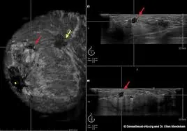
A breast ultrasound is a diagnostic imaging test that provides real-time images of breast tissues, including skin, fat, connective tissue, and milk ducts. It uses high-frequency sound waves to create images of the internal structures of the breast. This method is particularly useful in differentiating solid masses from fluid-filled cysts.

What Can Breast Ultrasound Show?
- Lumps and Masses: Helps detect both benign and malignant masses.
- Cysts: Can distinguish fluid-filled cysts from solid lumps.
- Tissue Density: Provides information on tissue density and structure.
- Abnormalities: Detects changes in breast tissue that might indicate tumors.
- Guidance for Procedures: Used to guide biopsies and needle aspirations.
- Blood Flow: Assesses the blood flow within masses to help differentiate between benign and malignant lesions.
Why Might You Need a Breast Ultrasound?
- Lump Detection: To evaluate any lumps or thickening found during a physical exam or mammogram.
- Young Women: Often used for women under 30, where mammograms are less effective due to denser breast tissue.
- Pregnancy: Safe to use during pregnancy when X-ray based imaging isn't recommended.
- Breast Pain: Helps to diagnose the causes of localized breast pain or discomfort.
- Follow-up: Used to monitor known breast conditions or response to treatments.
- Dense Breasts: Improves detection of abnormalities in women with dense breasts.
How is a Breast Ultrasound Performed?
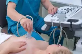
A breast ultrasound is a straightforward and painless procedure that typically takes about 20 to 30 minutes. Here's an overview of what to expect:
- Preparation: You'll be asked to remove any clothing and jewelry from the waist up. You might be given a gown to wear.
- Positioning: You will lie on your back on an examination table, and you might need to raise your arm above your head on the side being examined. This position helps spread the breast tissue evenly for better imaging.
- Gel Application: A clear, water-based gel is applied to the skin of the breast. This gel helps the transducer (the handheld device) make good contact with your skin, which is essential for producing clear images.
- Image Acquisition: The ultrasound technician or radiologist will move the transducer across your breast, taking images from different angles. The transducer emits high-frequency sound waves that penetrate the tissues and bounce back to create images on a monitor.
- Real-Time Viewing: The images are viewed on a screen in real-time, allowing the technician or radiologist to examine different areas of the breast. They might also take measurements of any identified masses or areas of interest.

- Completion: Once the images have been acquired, the gel will be wiped off your skin. There are usually no restrictions on activity after the procedure and you can resume normal activities immediately.
How to Interpret Breast Ultrasound Results
Understanding your ultrasound results can help you manage your health effectively. Here’s how you can approach interpreting your breast ultrasound results.
1. Utilizing X-ray Interpreter
X-ray Interpreter now provides AI-driven analysis for ultrasound images. Here's how to use it:
- Registration: Sign up on X-ray Interpreter to use our AI for ultrasound analysis.
- Uploading Ultrasounds: Upload your breast ultrasound images.
- Reviewing Interpretation: Receive an AI-generated interpretation, including a report.
- Consultation: Always consult with your physician for a complete diagnosis.
Check our get started guide for more details.
2. Using ChatGPT Plus
ChatGPT Plus can assist in analyzing ultrasound images through its advanced GPT-4V model:
- Subscription: Subscribe to ChatGPT Plus for access.
- Uploading Ultrasounds: Upload your ultrasound images on the OpenAI platform.
- Request Analysis: Ask the AI to interpret your ultrasound images.
- Review and Validate: Verify the AI's interpretation with a medical professional.
Find out more in our blog on using ChatGPT Plus for medical image interpretation.
Alternatively, as several other AI models with vision capabilities emerge, you can also try other models, such as Grok by xAI, Claude by Anthropic, Gemini by Google Deepmind.
3. Understanding the Basics Yourself
While not a substitute for professional advice, basic understanding can aid in communication with your doctor.
- Learn Anatomy: Understand the basic structure of the breast and common findings in ultrasounds.
- Review Guides: Consult reliable sources to learn about common breast abnormalities.
- Ask Questions: Note any unfamiliar terms and discuss them with your healthcare provider.
- Professional Validation: Always validate your understanding with a medical professional.
Comparing the Different Approaches
Let's compare the different methods for interpreting breast ultrasounds:
| Criteria | X-ray Interpreter | ChatGPT Plus | Self-Reading |
|---|---|---|---|
| Accuracy | High (AI-based)1 | High (AI-based)1 | Varies (Skill-dependent) |
| Ease of Use | Easy | Moderate | Challenging |
| Cost | Starting from $2.50 per image | $20 per month | Free (excluding educational costs) |
| Time Efficiency | Fast | Moderate to Fast | Slow to Moderate |
| Learning Curve | Low | Low to Moderate | High |
| Additional Resources | Provided | Partially Provided (through OpenAI) | Self-sourced |
Each method provides value with its unique strengths and weaknesses. AI-driven options offer fast and precise results, while basic knowledge enhances communication and self-awareness.
Conclusion
Breast ultrasound is an essential diagnostic tool for evaluating breast health, aiding in the identification of various breast abnormalities. This guide has provided insights into breast ultrasound technology and how to approach understanding your results with AI tools and self-guided research.
Always consider your specific needs and preferences when choosing an approach. Be mindful of data privacy and seek validation from qualified healthcare professionals.