How to Interpret Breast CT Scans: 3 Essential Methods
Breast CT scans are a powerful imaging technique used to visualize the breast tissue. This guide explains what a breast CT scan can reveal, why it's used, and how you can interpret its findings effectively.
What is a Breast CT Scan?
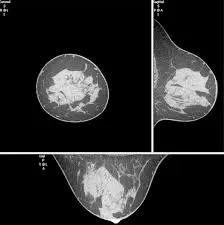
A breast CT (Computed Tomography) scan is an advanced imaging test that uses X-rays to create detailed cross-sectional images of the breast tissue. Unlike a mammogram, a CT scan provides 3D images, giving a more thorough view of the breast and surrounding structures. Let's explore how this technique works and what it can reveal.

What Can Breast CT Scans Show?
- Detailed Tissue Imaging: High-resolution images of breast tissue, including fat, glands, and connective tissues.
- Detection of Abnormalities: Identify masses, tumors, cysts, and calcifications, both benign and malignant.
- Lymph Node Evaluation: Clear visualization of the axillary lymph nodes, assisting in staging of breast cancer.
- Assessment of Chest Wall: Images of the chest wall and surrounding structures can be obtained.
- Monitoring Treatment Response: Used to monitor the effectiveness of cancer treatments.
Why Might You Need a Breast CT Scan?
- Suspicious Mammogram Findings: Used to investigate suspicious results from a mammogram or other breast imaging.
- Staging Breast Cancer: To determine the extent of breast cancer and involvement of lymph nodes.
- Monitoring Cancer Progress: To check the response of tumors to treatment.
- Evaluating Chest Pain: To rule out breast conditions as a cause of chest pain.
- Planning for Surgery or Therapy: To help surgeons and radiation oncologists with planning.
How is a Breast CT Scan Performed?
A breast CT scan is a non-invasive procedure that involves lying on a table while a scanner takes images of your breast tissue. Here is an overview of the process:

Step-by-Step Process:
- Preparation: You will be asked to remove any jewelry or metal objects that might interfere with the scan. You may be asked to wear a gown.
- Positioning: You'll lie on a specialized table that moves inside the CT scanner. You may be asked to raise your arms above your head to ensure clear imaging of the breast area.
- Scanning: The scanner uses X-rays to capture detailed cross-sectional images of your breasts. The process is quick, generally taking just a few minutes.
- Contrast (Sometimes): In some cases, a contrast dye may be administered intravenously to enhance the images. This allows better visualization of blood vessels and certain types of tissue.
- Completion: Once the scanning process is complete, the table will move back out of the scanner, and you will be free to go about your day. There are no post-procedure restrictions, unless a contrast dye is used.
The entire procedure is typically painless and doesn't require sedation. If a contrast dye was used, you might be asked to drink extra fluids to help flush it out of your system.
How to Interpret Breast CT Scan Results
Understanding the results of your breast CT scan is vital. Here's a rundown on how you can approach interpreting the findings.
1. Utilizing X-ray Interpreter
X-ray Interpreter now provides AI-driven analysis for CT scan images. Here's how you can use it:
- Registration: Sign up on X-ray Interpreter to begin analyzing your CT scans with AI.
- Uploading Images: Upload your breast CT scan images through our platform.
- Reviewing Interpretation: Receive an AI-generated report with interpretative analysis.
- Consultation: Always consult with your physician for a comprehensive diagnosis.
Check out our get started guide for more details.
2. Using ChatGPT Plus
ChatGPT Plus, enhanced with the GPT-4V model, can also assist with interpreting CT scans:
- Subscription: Subscribe to ChatGPT Plus to use its advanced features.
- Uploading Images: Upload your breast CT images onto the OpenAI platform.
- Request Analysis: Instruct the AI to analyze the scan and generate a report.
- Review and Validate: Always review AI-generated reports and validate them with a healthcare professional.
Find out more in our blog on using ChatGPT Plus for medical image interpretation.
Alternatively, as several other AI models with vision capabilities emerge, you can also try other models, such as Grok by xAI, Claude by Anthropic, Gemini by Google Deepmind.
3. Understanding the Basics Yourself
While not a replacement for medical professionals, understanding some basic concepts can help you participate in discussions with your doctor more effectively:
- Basic Anatomy: Learn about the basic anatomy of the breast, including lobes, ducts, and lymph nodes.
- Online Resources: Use reputable websites to learn about common findings in breast CT scans.
- Medical Terminology: Familiarize yourself with basic medical terminology related to breast imaging.
- Validate with Experts: Always ensure your understanding aligns with what your healthcare provider says.
Comparing the Different Approaches
Let’s compare different methods to analyze your breast CT scan results:
| Criteria | X-ray Interpreter | ChatGPT Plus | Self-Reading |
|---|---|---|---|
| Accuracy | High (AI-based)1 | High (AI-based)1 | Varies (Skill-dependent) |
| Ease of Use | Easy | Moderate | Challenging |
| Cost | Starting from $2.50 per image | $20 per month | Free (excluding educational costs) |
| Time Efficiency | Fast | Moderate to Fast | Slow to Moderate |
| Learning Curve | Low | Low to Moderate | High |
| Additional Resources | Provided | Partially Provided (through OpenAI) | Self-sourced |
Each method offers different benefits. AI-based tools offer quick and precise analyses, while basic understanding facilitates communication with medical professionals.
Conclusion
Breast CT scans are a critical tool in the diagnosis and management of breast conditions. This guide has detailed how these scans are used, what they show, and the methods available to help you understand the results.
Choosing a method depends on your specific needs, comfort level, and the resources at your disposal. Always ensure your results are validated by a healthcare expert to make informed decisions about your health.
Related Articles
- How To Interpret Chest CT Scans
- How To Interpret Mammograms
- How To Interpret Breast Ultrasounds
- How To Interpret Breast MRIs