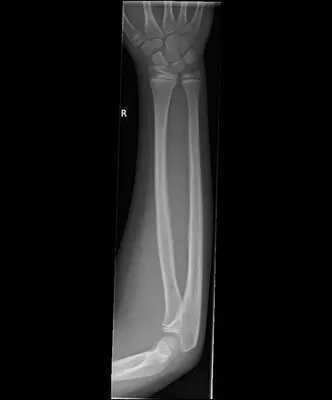
How to interpret arm X-rays: 3 Essential Techniques
Introduction
Mastering the art of arm X-ray analysis is crucial for orthopedic specialists, radiologists, and medical students. It provides vital insights for accurate diagnosis, effective treatment strategies, and holistic patient management. This guide aims to familiarize practitioners and learners with the key aspects of interpreting arm radiographs, with a special focus on the different types of X-rays involved. Our guide, designed for both experienced professionals and novices, breaks down intricate concepts into easy-to-understand steps, ensuring a comprehensive understanding of this essential diagnostic tool.
Understanding Arm X-rays: A Comprehensive Guide on Various Types
Arm X-rays are indispensable radiographic tools in orthopedics and trauma medicine. These imaging techniques offer detailed views of bones, joints, and soft tissues in the arm, from the shoulder to the wrist. This section aims to elucidate what arm X-rays are and their critical role in orthopedic diagnostics. By comprehending the unique features of different arm X-ray types, medical professionals can make better-informed decisions in various clinical situations, from fracture analysis to joint disorder evaluations.
Different Types of Arm X-rays: Understanding Their Uses
Arm X-rays can be categorized into several types, each serving a unique purpose in the evaluation of upper limb health.
Plain Radiographs: These are standard X-rays that provide two-dimensional images of the arm, useful for assessing fractures, dislocations, and bone diseases.
Fluoroscopy: A real-time arm imaging technique, perfect for guiding procedures like joint injections or assessing dynamic bone or joint issues.
CT Scans: Offer detailed, cross-sectional images of the arm, crucial for complex fracture assessments and surgical planning.
MRI: Provides detailed images of soft tissues, such as muscles, tendons, and ligaments, along with bones and joints, ideal for diagnosing soft tissue injuries and joint disorders.

Indications for Arm X-rays: When and Why They Are Essential
Arm X-rays are critical diagnostic tools in specific orthopedic conditions and scenarios. Key reasons for obtaining an arm X-ray include:
- Trauma and Injury: Essential for evaluating suspected fractures, dislocations, and other traumatic injuries.
- Chronic Pain or Dysfunction: Helps in diagnosing chronic conditions like arthritis or tendonitis.
- Preoperative Planning: Crucial for planning surgical procedures on the arm.
- Postoperative Evaluation: Used for assessing the success of surgical interventions and monitoring healing.
Recognizing when and why arm X-rays are necessary is vital for medical professionals and patients in managing upper limb health effectively.
Expert Techniques for Interpreting Arm X-rays
Interpreting arm X-rays requires skill and the right tools. Here are three effective methods:
1. AI-Assisted Analysis with X-ray Interpreter
X-ray Interpreter employs advanced AI technology for rapid, accurate interpretations of arm X-ray images. It's intuitive and ideal for dependable analysis.
To use X-ray Interpreter for AI-generated arm X-ray analysis, follow these steps:
- Registration: Sign up on X-ray Interpreter to access the AI-based analysis.
- Uploading X-rays: Upload your arm X-ray images onto the platform.
- Reviewing Interpretation: Examine the AI-generated interpretation and download the report.
- Seeking Professional Advice: Consult with medical professionals for further understanding of the AI interpretation.
Please check out our get started guide.
2. Interactive Analysis with ChatGPT Plus
ChatGPT Plus uses the powerful GPT-4V model to analyze arm X-ray images. This method offers an interactive experience, allowing dialogue with the AI to refine the analysis:
- Subscription: Subscribe to ChatGPT Plus for GPT-4V image analysis.
- Uploading X-rays: Go to the GPT-4V interface on the OpenAI platform and upload your arm X-ray images.
- Requesting Analysis: Ask for an analysis of the X-ray images with natural language commands or queries.
- Reviewing and Confirming Analysis: Examine the provided analysis, iterating for more detail if needed.
- Consulting Professionals: Seek validation of the analysis from medical professionals.
Please read our post on how to use ChatGPT Plus for arm X-ray analysis.
Alternatively, as several other AI models with vision capabilities emerge, you can also try other models, such as Grok by xAI, Claude by Anthropic, Gemini by Google Deepmind.
3. Self-Reading Arm X-rays
Self-reading is a traditional method reliant on individual expertise. Ideal for professionals aiming to improve their interpretive skills, this method demands a solid base in radiographic knowledge:
- Education: Gain foundational knowledge and training in arm X-ray interpretation from credible sources or courses.
- Practice: Regularly practice interpreting arm X-rays under expert supervision.
- Resources: Use books, online materials, and medical literature to enhance your interpretive skills.
- Seeking Feedback: Obtain critiques from experienced professionals to refine your interpretations.
- Continuous Learning: Constantly update your skills by engaging in medical literature, attending workshops, and participating in professional discussions.
Recommended Resources for Self-Reading:
Recommended Resources for Self-Reading:
-
X-ray Interpretation: Upper Limb Injuries by Radiopaedia.org:
- This course teaches radiographic interpretation with a focus on upper limb injuries. It includes audio and video commentary, over 300 annotated cases, and offers a completion certificate. Ideal for a broad audience including doctors, medical students, and healthcare professionals involved in the care of upper limb injuries.
-
X-Ray Exam: Upper Arm (Humerus) for Parents by KidsHealth:
- Provides information on what a humerus X-ray is, how it's performed, and why it's done. It details the procedure and the types of views taken during the exam, helping in understanding the process and results of upper arm X-rays.
-
X-Ray Exam: Forearm for Parents by KidsHealth:
- Explains what a forearm X-ray is, the procedure involved, and its purposes. This resource is valuable for parents and practitioners to understand forearm X-rays, including how to prepare and what to expect during the procedure.
-
Upper Extremity X-Ray by Cedars-Sinai:
- Details the process and purpose of upper extremity X-rays, which may include the fingers, hands, wrists, elbows, forearms, upper arms, or shoulders. It describes the procedure, including patient positioning and the duration of the X-ray.
Comparative Review of X-ray Interpretation Methods
Choosing the right method for interpreting arm X-rays is crucial for precise diagnosis and patient care. In this comparative analysis, we assess the three methods - AI-assisted interpretation with X-ray Interpreter, interactive analysis with ChatGPT Plus, and traditional self-reading. Our comparison focuses on accuracy, ease of use, cost, efficiency, and available resources. This analysis aims to give a clear understanding of each method's strengths and weaknesses, aiding in informed decision-making for arm X-ray interpretation.
| Criteria | X-ray Interpreter | ChatGPT Plus | Self-Reading |
|---|---|---|---|
| Accuracy | Mostly High (AI-based)1 | Mostly High (AI-based)1 | Varies (Skill-dependent) |
| Ease of Use | Easy | Moderate | Challenging |
| Cost | Starting from $2.50 per image | $20 per month | Free (excluding educational costs) |
| Time Efficiency | Fast | Moderate to Fast | Slow to Moderate |
| Learning Curve | Low | Low to Moderate | High |
| Additional Resources | Provided | Partially Provided (through OpenAI) | Self-sourced |
Conclusion: The Significance of Accurate Arm X-ray Analysis
Accurate interpretation of arm X-rays is a key skill in the medical field, crucial for diagnosing and managing various upper limb conditions. This guide offers an overview of three different methods to interpret arm X-rays: using X-ray Interpreter, ChatGPT Plus, and self-reading. Each method serves different expertise levels and situations, presenting a spectrum of options for individuals and professionals.
In the ever-evolving field of medical diagnostics, staying current with new technologies and methods is essential for providing accurate and timely patient care. Regardless of the chosen method, adherence to legal and ethical standards is vital for ensuring patient privacy, safety, and well-being.
This guide is a starting point for exploring diverse methods of arm X-ray interpretation, aiming to assist individuals and professionals in this critical aspect of medical diagnostics. The choice among these methods depends on personal preferences, professional objectives, and the specific context at hand.
Related Articles
Resources and Further Learning
For those interested in further exploring the field of arm X-ray interpretation and related medical imaging, several informative online resources are available:
-
Extremity X-Ray by Kaiser Permanente: This resource provides an overview of extremity X-rays, including their purpose and the types of conditions they can help diagnose, such as fractures, dislocations, infections, arthritis, bone growths, and other bone diseases.
-
X-rays - What Patients Need to Know by IAEA: This informative page by the International Atomic Energy Agency offers insights into the nature of X-rays, their medical applications, and safety considerations. It explains the uses of different types of X-ray-based procedures such as radiography, fluoroscopy, and computed tomography, as well as discussing the rare instances of adverse effects from radiation doses in diagnostic practice.
- Offers detailed information on forearm X-rays, including the standard projections used, indications for the exam, and how the X-ray series examines the radius and ulna. Useful for understanding the diagnostic process of forearm injuries.