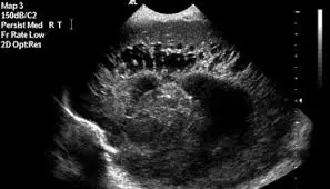
How to interpret Neonatal Cranial Ultrasound: 3 Key Aspects
Neonatal cranial ultrasound is a crucial imaging technique for examining a newborn's brain. This guide explains what it reveals, its purpose, and how to understand its findings.
What is Neonatal Cranial Ultrasound?
Neonatal cranial ultrasound is a non-invasive, real-time imaging test that uses sound waves to examine a newborn's brain. It's safe, painless, and vital for detecting various brain conditions. Let's explore this technique and its significance.

What Can Neonatal Cranial Ultrasound Show?
- Brain Structures: Clear visualization of the brain's ventricles, parenchyma, and other key structures.
- Hemorrhages: Detection of intraventricular hemorrhages (IVH) or other bleeding within the brain.
- Cysts and Lesions: Identification of cysts, masses, or other abnormalities.
- Hydrocephalus: Assessment for the presence of excess fluid in the brain (hydrocephalus).
- Real-time Imaging: Enables assessment of cerebral blood flow and dynamic changes.
Why Might a Neonate Need a Cranial Ultrasound?
- Prematurity: To assess for complications common in premature infants, such as IVH.
- Birth Complications: To evaluate for potential brain injuries sustained during delivery.
- Suspected Infections: To look for signs of brain infections or inflammation.
- Neurological Symptoms: To investigate seizures, abnormal reflexes, or other neurological issues.
- Monitoring: To follow up on known brain conditions and assess treatment response.
How a Neonatal Cranial Ultrasound is Performed
The procedure for a neonatal cranial ultrasound is straightforward, focusing on safety and comfort for the newborn. Here's a step-by-step overview:
-
Preparation: The baby is usually placed in a supine position (on their back), often in a warmer or incubator to maintain their body temperature. A small amount of ultrasound gel is applied to the anterior fontanelle (the soft spot on top of the baby’s head) or the temporal region.
-
Probe Placement: A specially designed ultrasound transducer (probe) is gently placed on the baby's head over the gel. The anterior fontanelle provides a good acoustic window to view the brain without interference from the skull bones.
-
Image Acquisition: The clinician moves the transducer across the fontanelle, capturing images of different parts of the brain. Real-time images are displayed on the ultrasound machine's monitor, allowing for immediate assessment.
-
Scanning Planes: The clinician obtains images in multiple planes (coronal, sagittal, and sometimes axial) to visualize the brain from different angles and ensure comprehensive coverage.
-
Evaluation and Interpretation: The sonographer or radiologist examines the images to identify any abnormalities, measure ventricles, and assess the overall brain structure.
-
Duration: The entire procedure typically takes about 15-30 minutes, depending on the complexity of the case and the number of images needed. It is non-invasive and does not involve any radiation.

How to Interpret Neonatal Cranial Ultrasound Results
Interpreting cranial ultrasound results is crucial for managing neonatal health. Here's how to approach it using different methods.
1. Utilizing X-ray Interpreter
X-ray Interpreter offers AI-driven analysis of neonatal cranial ultrasounds:
- Registration: Sign up on X-ray Interpreter to access AI ultrasound analysis.
- Uploading Ultrasounds: Upload your neonatal cranial ultrasound images.
- Reviewing Interpretation: Receive the AI-generated report with key findings.
- Consultation: Always discuss the results with a healthcare professional.
Check our get started guide for more details.
2. Using ChatGPT Plus
ChatGPT Plus, powered by GPT-4V, can also help analyze cranial ultrasounds:
- Subscription: Subscribe to ChatGPT Plus for advanced analysis.
- Uploading Ultrasounds: Upload your ultrasound images on the OpenAI platform.
- Request Analysis: Ask the AI to interpret the images and provide a report.
- Review and Validate: Confirm the AI's interpretation with a healthcare provider.
Find out more in our blog on using ChatGPT Plus.
Alternatively, as several other AI models with vision capabilities emerge, you can also try other models, such as Grok by xAI, Claude by Anthropic, Gemini by Google Deepmind.
3. Understanding the Basics Yourself
While not a substitute for professional guidance, learning the basics can aid understanding.
- Learn Anatomy: Familiarize yourself with the basic structures of a newborn's brain.
- Review Simple Guides: Utilize reliable sources to understand common ultrasound findings.
- Prepare Questions: Note any unusual terms to ask your healthcare provider.
- Seek Expert Opinion: Always validate your understanding with a medical expert.
Comparing the Different Approaches
Let's compare different methods for interpreting neonatal cranial ultrasounds:
| Criteria | X-ray Interpreter | ChatGPT Plus | Self-Reading |
|---|---|---|---|
| Accuracy | High (AI-based)1 | High (AI-based)1 | Varies (Skill-dependent) |
| Ease of Use | Easy | Moderate | Challenging |
| Cost | Starting from $2.50 per image | $20 per month | Free (excluding educational costs) |
| Time Efficiency | Fast | Moderate to Fast | Slow to Moderate |
| Learning Curve | Low | Low to Moderate | High |
| Additional Resources | Provided | Partially Provided (through OpenAI) | Self-sourced |
Each method has its own benefits. AI-based tools provide quick and precise results, while basic knowledge improves patient communication.
Conclusion
Neonatal cranial ultrasound is an essential tool for assessing a newborn's brain health. This guide explains the procedure, its significance, and methods to better understand the findings using AI and self-learning resources.
Consider your needs, level of understanding, and available resources when selecting a method. Always maintain privacy standards and seek professional medical validation.