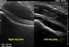
How to Interpret Joint Ultrasounds: 3 Essential Methods
Joint ultrasound is a vital imaging technique used to evaluate joints and surrounding tissues. This guide will explore what a joint ultrasound can show, why it's used, and how you can approach interpreting the results.
What is Joint Ultrasound?
Joint ultrasound is a non-invasive imaging method that utilizes sound waves to create real-time images of joints, ligaments, tendons, and soft tissues. It's safe, painless, and doesn't use radiation. Let’s understand what this technique involves and what it can show.

What Can Joint Ultrasound Show?
- Joint Effusions: Detects abnormal fluid accumulation within the joint space.
- Tendon Issues: Visualizes tendinitis, tears, or thickening of tendons around the joints.
- Ligament Injuries: Helps identify sprains, tears, and inflammation of ligaments.
- Cartilage Assessment: Provides limited assessment of cartilage integrity.
- Soft Tissue Abnormalities: Identifies masses, cysts, or inflammation in the soft tissues.
- Bone Surface Irregularities: Can show surface changes and bone outgrowths.
- Real-time Assessment: Allows for dynamic imaging during joint movement.
Why Might You Need a Joint Ultrasound?
- Joint Pain: To evaluate the cause of unexplained joint discomfort.
- Swelling: To assess for joint effusions and soft tissue inflammation.
- Limited Range of Motion: To help identify the source of movement restrictions.
- Sports Injuries: To diagnose tendon, ligament, and muscle related injuries.
- Follow-Up Monitoring: To check the progress of known joint conditions.
- Guided Procedures: To guide needle placement for joint injections or aspirations.
How is a Joint Ultrasound Performed?
A joint ultrasound is a quick and painless procedure. Here's what you can expect:
- Preparation: You may be asked to remove any jewelry or clothing covering the area to be examined.
- Positioning: You'll be positioned comfortably, either sitting or lying down, depending on the joint being scanned.
- Gel Application: A clear, water-based gel will be applied to the skin over the joint. This gel helps the sound waves travel effectively.
- Scanning: A transducer, which looks like a small microphone, will be moved over the gelled area. This transducer emits sound waves and receives the echoes, creating real-time images on a monitor.
- Image Review: The sonographer or doctor will review the images to assess the joint and surrounding tissues for any abnormalities.
- Clean Up: The gel will be wiped off, and you can return to your activities immediately after the scan.

How to Interpret Joint Ultrasound Results
Interpreting a joint ultrasound involves understanding normal anatomy and recognizing pathological changes. Here are three methods to approach interpretation.
1. Utilizing X-ray Interpreter
X-ray Interpreter provides AI-driven analysis to assist with ultrasound interpretation:
- Registration: Sign up on X-ray Interpreter to use our AI for ultrasound analysis.
- Uploading Images: Upload your joint ultrasound images to the platform.
- Reviewing Interpretation: Receive the AI-generated interpretation report.
- Consultation: Always discuss the results with your physician.
Check our get started guide for more details.
2. Using ChatGPT Plus
ChatGPT Plus, with its GPT-4V model, can also assist in analyzing ultrasound images:
- Subscription: Subscribe to ChatGPT Plus for advanced analysis.
- Uploading Images: Upload your ultrasound images to the OpenAI platform.
- Requesting Analysis: Ask the AI to interpret the ultrasound images.
- Review and Validate: Review the results, always confirming with a healthcare professional.
Find out more in our blog on using ChatGPT Plus for medical image interpretation.
Alternatively, as several other AI models with vision capabilities emerge, you can also try other models, such as Grok by xAI, Claude by Anthropic, Gemini by Google Deepmind.
3. Understanding the Basics Yourself
While not a substitute for medical professionals, some basic understanding can help you better understand your results.
- Learn Anatomy: Familiarize yourself with basic joint anatomy.
- Review Simple Guides: Utilize online resources to learn about common findings in joint ultrasounds.
- Note Questions: Note down unfamiliar terms and ask your healthcare provider.
- Seek Expert Guidance: Always validate your understanding with a medical professional.
Comparing the Different Approaches
Here’s a comparison of the different methods for interpreting joint ultrasounds:
| Criteria | X-ray Interpreter | ChatGPT Plus | Self-Reading |
|---|---|---|---|
| Accuracy | High (AI-based)1 | High (AI-based)1 | Varies (Skill-dependent) |
| Ease of Use | Easy | Moderate | Challenging |
| Cost | Starting from $2.50 per image | $20 per month | Free (excluding educational costs) |
| Time Efficiency | Fast | Moderate to Fast | Slow to Moderate |
| Learning Curve | Low | Low to Moderate | High |
| Additional Resources | Provided | Partially Provided (through OpenAI) | Self-sourced |
Each method offers advantages and disadvantages. AI options are fast and precise, while a basic understanding enables more informed patient-doctor communication.
Conclusion
Joint ultrasounds are a valuable tool for diagnosing a variety of musculoskeletal conditions. This guide has introduced you to the use of joint ultrasound technology, the images they produce, and ways you can better understand your results.
When choosing a method, consider your needs, level of understanding, and available resources. Always be aware of privacy standards and seek expert medical validation.