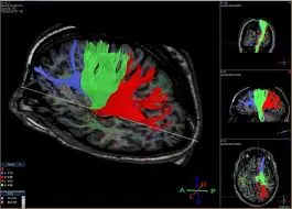
How to Interpret Diffusion Tensor Imaging: 3 Essential Techniques
Understanding Diffusion Tensor Imaging (DTI) is crucial for diagnosing and treating neurological conditions effectively. This guide introduces you to the DTI's advantages and explains three key techniques for interpreting these advanced images.
Understanding Diffusion Tensor Imaging
Diffusion Tensor Imaging provides a comprehensive view of neural pathways, offering detailed images of white matter tracts and brain connectivity. This section outlines what DTI is and its advantages for diagnosing various conditions.

What Does Diffusion Tensor Imaging Show?
- White Matter Tracts: Superior imaging of brain's white matter pathways.
- Brain Connectivity: Clear visualization of neural connections, useful in diagnosing connectivity issues.
- Microstructural Changes: Detects changes within white matter not visible on conventional MRIs.
- Disease Detection: More effective in identifying conditions like multiple sclerosis and traumatic brain injuries.
When Will You Get One?
- Persistent Neurological Symptoms: Especially if not adequately explained by conventional MRI.
- Neurodegenerative Diseases: Such as Alzheimer's disease or multiple sclerosis.
- Pre-surgical Planning: Provides a detailed map for surgical planning in brain surgeries.
- Research: Ideal for studying brain connectivity and microstructural changes in research settings.
Techniques to Interpret Diffusion Tensor Imaging
Interpreting DTI involves a deeper understanding of advanced imaging techniques. Here are three techniques for professionals at different levels of expertise.
1. Utilizing X-ray Interpreter
X-ray Interpreter now extends its AI-driven analysis to DTI images. The process ensures precision:
- Registration: Register on X-ray Interpreter to access AI analysis for DTI.
- Uploading DTIs: Upload your DTI images.
- Reviewing Interpretation: Receive the AI-generated interpretation and download your report.
- Professional Consultation: Always advisable to consult with medical professionals for comprehensive understanding.
Please check out our get started guide.
2. Using ChatGPT Plus
ChatGPT Plus now supports DTI image analysis with its latest GPT-4V model, offering detailed and interactive insights:
- Subscription: Subscribe to ChatGPT Plus for advanced image analysis features.
- Uploading DTIs: Interface with GPT-4V on OpenAI to upload your DTI images.
- Requesting Analysis: Engage with the model for a thorough analysis.
- Review and Confirmation: Assess and refine the analysis as needed.
- Professional Validation: Validation by medical experts is recommended.
Read more on our post about using ChatGPT Plus for MRI interpretation.
Alternatively, as several other AI models with vision capabilities emerge, you can also try other models, such as Grok by xAI, Claude by Anthropic, Gemini by Google Deepmind.
3. Mastering DTI Interpretation Yourself
For healthcare professionals aiming to enhance their DTI reading skills, self-learning is invaluable:
- Education: Pursue advanced training in DTI interpretation.
- Practice: Regular practice under expert guidance.
- Resources: Utilize advanced imaging books and online courses.
- Feedback: Seek feedback to refine skills.
- Continuous Learning: Engage in ongoing education to stay current with imaging techniques.
Recommended Resources for Self-Learning:
-
Diffusion Tensor Imaging and Fibre Tractography - Radiopaedia: Provides a comprehensive overview of DTI and fibre tractography, including its applications, limitations, and the science behind the technique.
-
Diffusion Tensor Imaging - YouTube: A detailed video explaining the principles of DTI, its clinical applications, and how it is used to visualize neural pathways in the brain.
-
Diffusion Tensor Imaging - ScienceDirect: Offers in-depth articles and research papers on DTI, covering its methodology, clinical applications, and recent advancements in the field.
Comparative Analysis
Choosing the right technique for interpreting DTI is vital for accurate diagnosis. This section compares the three methods:
| Criteria | X-ray Interpreter | ChatGPT Plus | Self-Reading |
|---|---|---|---|
| Accuracy | High (AI-based)1 | High (AI-based)1 | Varies (Skill-dependent) |
| Ease of Use | Easy | Moderate | Challenging |
| Cost | Starting from $2.50 per image | $20 per month | Free (excluding educational costs) |
| Time Efficiency | Fast | Moderate to Fast | Slow to Moderate |
| Learning Curve | Low | Low to Moderate | High |
| Additional Resources | Provided | Partially Provided (through OpenAI) | Self-sourced |
Each method has its strengths and weaknesses, with AI options providing rapid and precise interpretations, while self-reading encourages in-depth learning for medical professionals.
Conclusion
DTI interpretation is critical for diagnosing complex neurological conditions. This guide presents three techniques suited to different professional needs and skill levels. AI methods offer quick, precise interpretations, while self-learning is geared towards those seeking in-depth knowledge.
When selecting a technique, consider your level of expertise, the need for timely interpretation, and the resources available. Ethical and legal standards must always be maintained to ensure patient safety and confidentiality.
Resources and Further Learning
For further exploration and understanding of DTI interpretation, consider the following resources:
-
Diffusion Tensor Imaging - Medscape: Provides an in-depth overview of DTI, including its clinical applications, techniques, and limitations.
-
Magnetic Resonance Imaging of the Brain - Wiley Online Library: An academic paper discussing various MRI techniques, including DTI, and their applications in medical imaging.
-
Functional MRI and Diffusion Tensor Imaging - Mayfield Clinic: An informative resource on the use of fMRI and DTI for visualizing brain activity and white matter pathways.