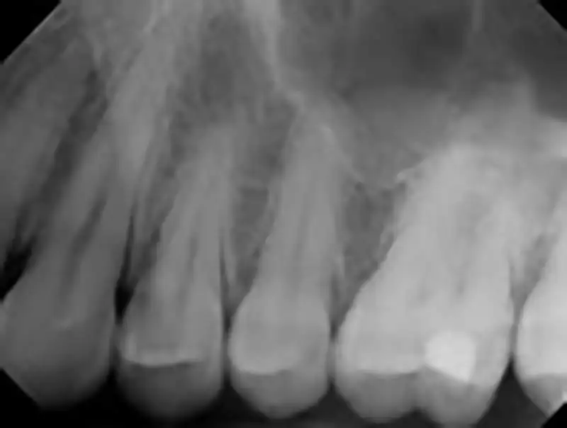
How to interpret dental X-rays: 3 Essential Methods
Introduction
Understanding the basics of dental X-ray analysis is a pivotal skill in both dentistry and orthodontics. It provides essential insights for accurate diagnosis, effective treatment planning, and comprehensive patient care. This guide is tailored to acquaint practitioners and students alike with the fundamentals of interpreting dental radiographs. Focusing particularly on Intraoral and Extraoral X-rays, we delve into the nuances of dental radiography interpretation. Whether you're a seasoned professional or a newcomer to the field, our step-by-step approach simplifies complex concepts, ensuring a thorough grasp of this critical diagnostic tool.
Exploring Dental X-rays: Detailed Guide on Intraoral and Extraoral Types
Dental X-rays, encompassing both Intraoral and Extraoral types, are crucial radiographic tools in modern dentistry. These imaging techniques capture detailed pictures of teeth, jawbones, and other structures within the oral cavity. In this section, we clarify what dental X-rays are and explore their indispensable role in dental diagnostics. By understanding the distinct features of Intraoral and Extraoral X-rays, including their applications and benefits, dental professionals can make informed decisions in various clinical scenarios, ranging from cavity detection to orthodontic assessments.
Intraoral vs. Extraoral X-rays: Understanding Different Types
Dental X-rays fall into two main categories: Intraoral and Extraoral. Each type serves a specific purpose in dental health assessment.
Intraoral X-rays: These are detail-oriented images focusing on individual teeth, their supporting bone structure, and surrounding tissues. They are pivotal for detecting cavities, monitoring tooth root health, and assessing the development of teeth. Common types include Bitewing X-rays, used for identifying tooth decay, and Periapical X-rays, which show the entire tooth from crown to root.

Extraoral X-rays: These provide a broader view of the entire oral cavity, including the jawbone and surrounding structures. They are essential for evaluating impacted teeth, examining jaw development, and identifying conditions like cysts or tumors. Panoramic X-rays, a type of Extraoral X-ray, are particularly useful for orthodontic planning and diagnosing emerging dental issues.

Indications for Dental X-rays: When and Why They Are Needed
Dental X-rays are not just routine procedures; they are essential tools for specific dental conditions and scenarios. The common reasons for requiring a dental X-ray include:
- Symptom Investigation: For persistent toothaches, jaw pain, or swelling, dental X-rays offer a clear view of underlying issues.
- Routine Dental Check-ups: Particularly crucial for individuals with a history of dental problems.
- Pre-Treatment Assessment: Essential before certain dental procedures to thoroughly assess oral health.
- Orthodontic Planning: Invaluable for evaluating teeth alignment and formulating precise orthodontic treatments.
Understanding when and why dental X-rays are necessary is key for both dental professionals and patients in managing oral health effectively.
Expert Techniques for Interpreting Dental X-rays
The interpretation of dental X-rays is a nuanced process, varying based on the expertise and tools at hand. We outline three effective methods:
1. AI-Assisted Analysis with X-ray Interpreter
X-ray Interpreter uses advanced AI technology for swift, accurate interpretations of dental X-ray images. It's user-friendly and ideal for quick, reliable analysis.
By following these steps, you can effortlessly obtain an AI-generated analysis of your X-ray images:
- Registration: Sign up on X-ray Interpreter to access the AI-based analysis.
- Uploading X-rays: Upload your dental X-ray images onto the platform.
- Reviewing Interpretation: Review the AI-generated interpretation and download the report.
- Seeking Professional Advice: If necessary, consult with dental professionals to understand the interpretation in a clinical context.
Please check out our get started guide.
2. Interactive Analysis with ChatGPT Plus
ChatGPT Plus leverages the potent GPT-4V model to provide insightful analysis of dental X-ray images. This method offers a more interactive experience, allowing you to communicate with the AI and refine the analysis to suit your needs:
- Subscription: Subscribe to ChatGPT Plus to access GPT-4V for image analysis.
- Uploading X-rays: Navigate to the GPT-4V interface on the OpenAI platform and upload your dental X-ray images.
- Requesting Analysis: Request an analysis of the X-ray images by inputting natural language commands or questions.
- Reviewing and Confirming Analysis: Review the analysis provided, iterate to get more detailed or specific information if necessary.
- Consulting Professionals: Seek advice from dental professionals to validate the analysis provided by GPT-4V.
Please read our post on how to use ChatGPT Plus for dental X-ray analysis to learn more.
Alternatively, as several other AI models with vision capabilities emerge, you can also try other models, such as Grok by xAI, Claude by Anthropic, Gemini by Google Deepmind.
3. Self-Reading Dental X-rays
Self-reading is a traditional method that relies on individual expertise and resources. It's ideal for dental professionals aiming to hone their interpretative skills. This method requires a strong foundation in dental knowledge and a willingness to continuously learn and improve:
- Education: Acquire basic knowledge and training on reading and interpreting dental X-rays from reputable sources or through coursework.
- Practice: Practice interpreting dental X-rays under the guidance of experienced professionals.
- Resources: Utilize books, online resources, and dental literature to enhance your understanding and skills in interpreting dental X-rays.
- Seeking Feedback: Obtain feedback from knowledgeable professionals to improve your interpretation skills.
- Continuous Learning: Continuously update your knowledge and skills by reading recent dental literature, attending workshops, and engaging in discussions with professionals.
Recommended Resources for Self-Reading:
-
How to Read Dental X-rays by DentQ International:
- A video guide explaining the basics of dental X-rays and how to interpret them, differentiating between various types of images and explaining typical radiolucency of structures like enamel, dentin, and bone, along with detecting various dental conditions using X-ray images.
-
Interpreting Dental X-rays: A Practical Guide for Dentists by Allied Academies:
- Emphasizes the importance of dental X-rays for diagnosing issues, planning treatments, and monitoring oral health, describing various types of dental X-rays and essential principles like diagnostic criteria, comparison with previous X-rays, and understanding radiographic anatomy.
-
How to Read X-rays by Desert Ridge Smiles:
- Provides a quick guide on interpreting dental X-rays, explaining how to differentiate between normal and diseased tooth and bone structures, showcasing how to identify areas of decay, and the implications of decay reaching different layers of a tooth.
Comparative Review of Dental X-ray Interpretation Methods
Choosing the right method for interpreting dental X-rays is crucial for accurate diagnosis and efficient patient care. In this comparative analysis, we evaluate the three methods - AI-assisted interpretation with X-ray Interpreter, interactive analysis with ChatGPT Plus, and traditional self-reading. Our comparison is based on accuracy, ease of use, cost, time efficiency, and the availability of additional resources. This evaluation aims to provide a clear understanding of each method's advantages and limitations, helping you make an informed decision tailored to your specific needs and expertise in dental X-ray interpretation.
| Criteria | DentalX-ray Interpreter | ChatGPT Plus | Self-Reading |
|---|---|---|---|
| Accuracy | Mostly High (AI-based)1 | Mostly High (AI-based)1 | Varies (Skill-dependent) |
| Ease of Use | Easy | Moderate | Challenging |
| Cost | Starting from $2.50 per image | $20 per month | Free (excluding educational costs) |
| Time Efficiency | Fast | Moderate to Fast | Slow to Moderate |
| Learning Curve | Low | Low to Moderate | High |
| Additional Resources | Provided | Partially Provided (through OpenAI) | Self-sourced |
Summing Up: The Importance of Accurate Dental X-ray Analysis
Interpreting dental X-rays is a crucial skill in the dental field, aiding in the diagnosis and management of numerous oral health conditions. This guide provided an overview of three different methods to interpret dental X-rays: utilizing DentalX-ray Interpreter, using ChatGPT Plus, and self-reading. Each method caters to different levels of expertise and circumstances, offering a range of options for individuals and professionals alike.
In a rapidly evolving dental landscape, staying updated with the latest technologies and methodologies is crucial for providing accurate and timely patient care. Regardless of the method chosen, adhering to legal guidelines and ethical practices is paramount to ensure the privacy, safety, and wellbeing of individuals.
This guide serves as a starting point in exploring the various methods of dental X-ray interpretation, with the hope of aiding individuals and professionals in navigating this crucial aspect of dental diagnostics. The choice between these methods ultimately depends on personal preferences, professional goals, and the specific circumstances at hand.
Related Articles
Resources and Further Learning
For those interested in delving deeper into the realm of dental X-ray interpretation, a plethora of resources is available online. These resources offer invaluable insights and structured approaches to interpreting dental X-rays, aiding in the enhancement of your skills and understanding in this critical aspect of dental diagnostics.
-
Dental X-Ray's Radiology Course on Udemy Discover the basics of dental anatomy and how it shows up on digital X-rays, covering different types of dental radiographic images. Ideal for individuals interested in pursuing dentistry, dental hygiene, or dental assisting.
-
Practical Panoramic Imaging - Dentalcare Course This course is designed to provide dental professionals with the knowledge to discuss radiographic selection criteria and the indications for panoramic imaging, compare and contrast panoramic and intraoral imaging, and understand the advantages and limitations of panoramic radiographic imaging.