Normal vs. Abnormal Chest X-Rays: What Do They Reveal?
Chest X-rays are not just pictures; they are windows into the health of your thoracic cavity, including lungs, heart, airways, blood vessels, and the bones of your spine and chest wall.
These detailed images are pivotal in distinguishing normal from concerning signs, steering clinicians toward accurate diagnoses and effective treatments.
Normal vs Abnormal Chest X-ray at a Glance
| Feature | Normal | Abnormal |
|---|---|---|
| Lung Fields | Clear, black (air-filled) | White patches, consolidation, collapse |
| Heart Size | <50% chest width (CTR < 0.5) | Enlarged (CTR > 0.5), possibly rounded |
| Diaphragm | Smooth, curved | Flattened (COPD), obscured (effusion) |
| Bones & Soft Tissues | Aligned, no masses | Fractures, masses, displaced structures |
Normal vs Abnormal Chest X-Ray: Key Differences
Looking to compare a normal chest X-ray vs abnormal one? This guide highlights exactly what to look for using real image examples, helping you better understand both expected structures and signs that something may be off.
What Does a Healthy Chest Look Like on an X-Ray?
What should you expect to see in a normal chest X-ray? Here are the visuals that suggest everything is working just fine.
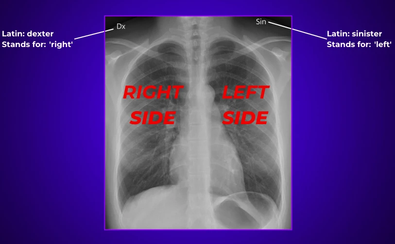
Firstly, when viewing a chest X-ray, it's important to remember that the left side of the image corresponds to the patient’s right side, and the right side of the image corresponds to the patient’s left side. This reversal helps align with the patient’s perspective.
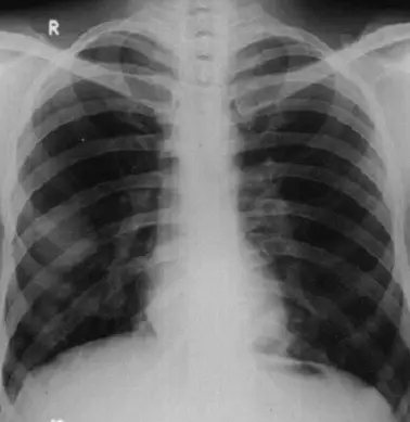
The lungs are divided into lobes, which are distinct sections separated by fissures:
- The right lung has three lobes: the upper lobe (RUL), middle lobe (RML), and lower lobe (RLL).
- The left lung has two lobes: the upper lobe (LUL) and lower lobe (LLL), with the space of the middle lobe taken up by the heart.

On a chest X-ray, these lobes are not always distinctly visible, but subtle differences in their density or markings can help identify abnormalities like consolidation, atelectasis, or fluid.
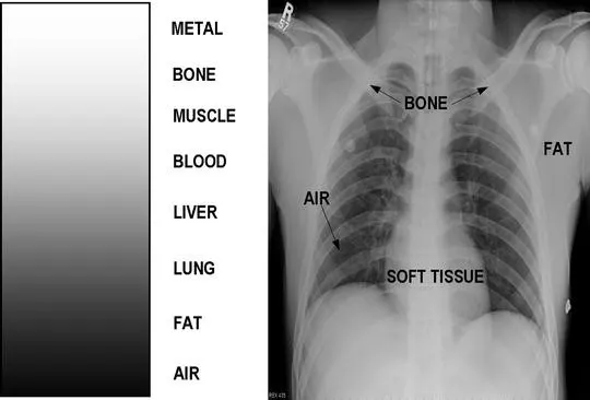
Lung Fields
Expect to see a lot of black, that's the air in your lungs, interlaced with delicate white lines that map out your airways and blood vessels.
Diaphragm and Heart Contours
Your diaphragm should show a neat, curved line, sitting higher on the right because of the liver underneath. The heart should not occupy more than half the chest width, its outline smooth and distinct.
Bone Integrity
Your ribs, spine, and clavicles should appear intact, aligned, and free from any fractures or unusual growths.

Soft Tissue Normalcy
The muscles and fat around your chest wall should look normal without any signs of swelling or masses.
What Can Go Wrong? Spotting Troubles in Chest X-Rays
When something's off, what abnormalities might your chest X-ray reveal? Let’s dive into the signs that call for a closer look, with each abnormality explained in more detail.
Are There Unwanted Shadows?
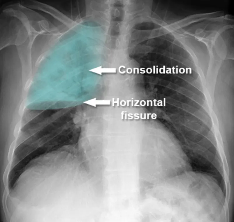
Shadows on a chest X-ray can be a red flag. These white patches,called opacities, might mean the lungs aren’t fully clear.
One common cause is consolidation, often seen with pneumonia. It looks like a dense, cloudy area in part of the lung, usually where infection or fluid has replaced air.
But not all shadows are created equal. Some may indicate benign nodules, scarring, or more serious concerns like lung cancer or tuberculosis. The size, shape, and location of the shadow help radiologists figure out what it could be.
If something suspicious is spotted, your doctor may follow up with a CT scan or recommend monitoring it over time.
Is There Excess Fluid?
Sometimes, the edges of the lungs or heart on an X-ray appear blurry or faded. This can be a clue that there’s fluid in the pleural space, the thin area between your lungs and chest wall.
This condition is called a pleural effusion, and it can make breathing harder or cause chest discomfort. It’s not a disease itself, but a symptom of something else going on.
Common causes include heart failure, kidney problems, liver disease, or infections like pneumonia or tuberculosis.
When fluid is suspected, doctors may order an ultrasound or CT scan to confirm it. Sometimes, they’ll drain the fluid using a procedure called thoracentesis to relieve pressure and find out what’s causing it.
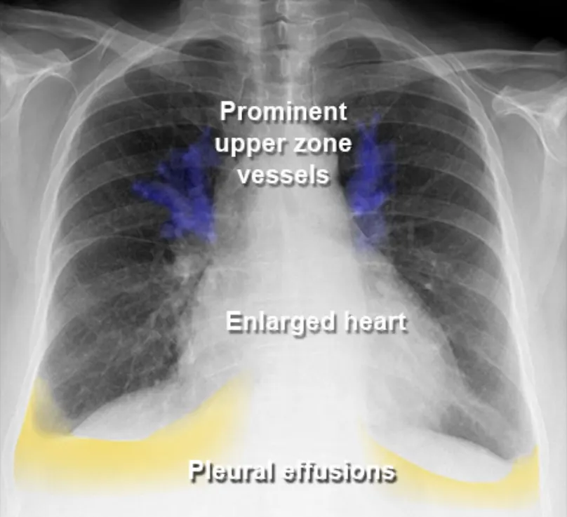
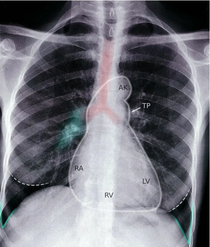
How Big is the Heart?
Your heart should take up less than half the width of your chest on a proper chest X-ray. When it looks bigger than that, doctors call it cardiomegaly, an enlarged heart.
This doesn’t always mean something serious, but it can be linked to conditions like high blood pressure, heart valve problems, or cardiomyopathy.
To measure it, radiologists use the Cardio-Thoracic Ratio (CTR), comparing the width of the heart to the total width of the chest. A CTR over 50% is usually considered abnormal.
If your heart appears enlarged, your doctor might suggest an echocardiogram or other heart tests to learn more.

CTR = Cardiac Width : Thoracic Width In this example, the CTR = 68%, which indicates enlargement.
To ensure accuracy, this measurement must be done on a Posterior-Anterior (PA) projection, as other angles can exaggerate heart size.
Are the Lungs Fully Inflated?
Lungs should look big, dark, and full of air on an X-ray. But sometimes, part of a lung collapses, a condition called atelectasis.
This shows up as a dense, white patch. The rest of the chest might try to compensate, and you may see the trachea or heart shift toward the collapsed side.
Atelectasis can happen after surgery, from mucus plugs, or due to tumors blocking airways. It might cause shortness of breath or go unnoticed if it’s small.
Follow-up imaging is often needed to figure out why it happened, and whether it resolves on its own or needs further treatment.

Is There Air Where It Shouldn’t Be?
There should never be air floating outside your lungs. If it gets into the pleural space, the gap between the lung and chest wall, it’s called a pneumothorax.
This looks like a clear space on an X-ray where lung markings suddenly disappear. It may also push the lung inward, making it shrink.
Pneumothorax can happen after injury, lung procedures, or even spontaneously in healthy people. Small ones might not need treatment, but larger ones can cause sharp chest pain and trouble breathing.
In those cases, doctors might insert a needle or chest tube to remove the air and let the lung re-expand.

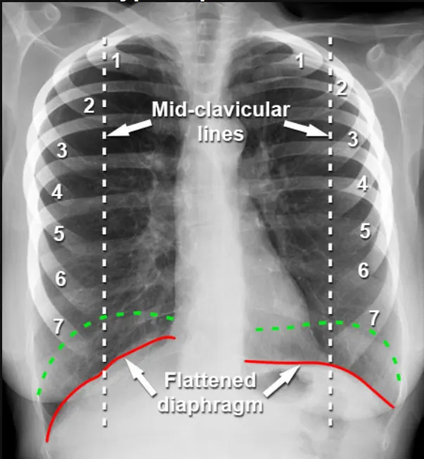
Do the Lungs Look Overly Large?
Lungs that look too big on an X-ray can be a sign of hyperinflation. This is common in people with chronic obstructive pulmonary disease (COPD).
When lungs are chronically overfilled with air, the diaphragm flattens and the chest may appear expanded. You might also see more space between the ribs and fewer visible blood vessels.
This happens because air gets trapped inside the lungs, making it harder to breathe out. It’s often linked to long-term smoking or other chronic lung conditions.
Treatment usually focuses on managing symptoms with inhalers, medications, lifestyle changes, and pulmonary rehab.
Are the Lymph Nodes Enlarged?
Lymph nodes don’t usually show up clearly on a chest X-ray. But when they do, and they look enlarged, it could mean something is wrong.
This finding is called hilar or mediastinal lymphadenopathy, and it may point to infections like tuberculosis, cancers like lymphoma, or autoimmune conditions such as sarcoidosis.
If enlarged nodes are seen, doctors may recommend a CT scan to get a better look. In some cases, a biopsy is needed to figure out what’s going on.

Visual Guide to Abnormal Chest X-Ray Findings

What’s Next? Understanding the Clinical Significance
What do these findings imply for further tests or treatments?
Chest X-rays are often the first step in identifying potential health concerns, but they don't always provide the complete picture. If your X-ray shows any abnormalities, such as consolidation, pleural effusion, cardiomegaly, atelectasis, pneumothorax, hyperinflated lungs, or enlarged lymph nodes, the next steps will depend on what was found and your overall health status.
Here's a breakdown of common abnormalities and what typically follows:
- Consolidation (e.g., pneumonia): May require blood tests, sputum cultures, and possibly a follow-up X-ray within a few days to monitor resolution.
- Pleural Effusion (fluid in the chest cavity): Often followed by ultrasound or CT scan to assess volume and cause. Thoracentesis may be performed to remove and test the fluid.
- Cardiomegaly (enlarged heart): Further evaluation with an echocardiogram, ECG (EKG), and blood tests for cardiac function.
- Atelectasis (lung collapse): May need repeat imaging, bronchoscopy, or pulmonary consultation, especially if recurrent or unexplained.
- Pneumothorax (air outside the lungs): Depending on size/severity, might need urgent intervention like a chest tube insertion or needle decompression. A CT scan may follow to rule out underlying causes.
- Hyperinflation (seen in COPD): Leads to pulmonary function testing, lifestyle evaluation, and possibly referral to a pulmonologist.
- Hilar or Mediastinal Lymphadenopathy: A CT scan or even biopsy may be needed to rule out infections, cancer, or autoimmune diseases.
In addition to imaging, doctors often consider your symptoms, medical history, and lab results to decide the next steps. Sometimes what appears abnormal may be incidental or age-related, while in other cases it might reveal a previously undetected condition that needs long-term management.
Early detection allows for early intervention, and follow-up testing is key to refining diagnosis and guiding treatment. So while an abnormal chest X-ray can be concerning, it also opens the door to taking action and getting answers.
How to Talk to Your Doctor? Questions to Ask After Your Chest X-Ray
It’s natural to have questions after receiving a chest X-ray report, especially if it mentions abnormalities. Clear communication with your healthcare provider is key to understanding what the results mean and what comes next.
Here are some helpful tips for your consultation:
- Ask for clarification: If a term like “infiltrate,” “opacity,” or “effusion” appears in your report, ask your doctor what it means in plain language.
- Discuss your symptoms: Let your doctor know if you’ve experienced cough, chest pain, shortness of breath, or recent infections.
- Request to see the image: Seeing the X-ray yourself (with your doctor’s explanation) can help you better understand the findings.
- Understand the timeline: Ask whether you need follow-up imaging or if it’s a one-time finding.
- Bring up your history: Past lung conditions, surgeries, or smoking history can change how results are interpreted.
By being open and engaged during your visit, you’ll gain clarity and make more informed decisions about your health.
What Comes After? Treatment Options Based on Chest X-Ray Findings
While a chest X-ray itself doesn’t determine treatment, it plays a key role in guiding the next steps. Treatments depend entirely on the underlying condition causing the abnormality.
Here are some common examples:
- Pneumonia or infection: Treated with antibiotics, rest, fluids, and possibly hospitalization in severe cases.
- Pleural effusion: May require fluid drainage and treatment of the underlying cause (e.g., heart failure, infection, cancer).
- Cardiomegaly: Often managed with medications for high blood pressure, heart failure, or underlying valve disease.
- Pneumothorax: Small cases may resolve on their own, while larger ones may need a chest tube or surgery.
- COPD/hyperinflation: Managed with inhalers, lifestyle changes (especially quitting smoking), and pulmonary rehab.
- Suspicious masses or lymphadenopathy: May lead to further imaging, biopsy, and treatment depending on the diagnosis (e.g., cancer, tuberculosis).
Every case is unique. A chest X-ray helps uncover the issue, but only a thorough clinical evaluation can determine the right course of action. If you’re ever unsure, don’t hesitate to ask for a second opinion or additional explanation. You can also explore our Radiology Center Directory to find a trusted imaging provider near you.
Are Chest X-Rays Safe? What You Should Know
Chest X-rays are one of the most common and trusted medical imaging tools, and they’re generally very safe. They use a small amount of ionizing radiation to create detailed images of the chest, including the lungs, heart, and bones.
The radiation dose from a standard chest X-ray is very low, about the same as what you’d be exposed to naturally from the environment over a few days.
Still, like all medical procedures, chest X-rays should be used thoughtfully. Doctors weigh the benefits against any minimal risk, especially for children and pregnant individuals. If you're unsure, it's always okay to ask your provider why the X-ray is being recommended.
What Can’t X-Rays Show? Knowing the Limitations
Chest X-rays are great for spotting many issues, but they do have limits.
They might miss very small tumors or early-stage infections. Some conditions, like blood clots in the lungs (pulmonary embolism), subtle heart valve problems, or early interstitial lung disease, aren’t clearly visible on a standard X-ray.
X-rays also provide only a flat, two-dimensional view. This means some overlapping structures can hide small issues.
That’s why doctors sometimes follow up with other imaging tests like CT scans, MRIs, or ultrasounds, depending on the case. Chest X-rays are a great starting point, but they’re rarely the final word.
Before you go...
How valuable is a chest X-ray? Extremely!
By accurately reading these images, healthcare providers can kickstart the most effective care plans, gaining crucial insights into your pulmonary, cardiac, and overall health.
Let's appreciate this fundamental diagnostic tool for the clarity it brings to medical evaluations.
If you're interested in further exploring the nuances of chest X-ray interpretation, why not try our X-ray Interpreter tool?
It's designed to assist both professionals and enthusiasts in understanding complex radiographic images.