Understanding the Anterior Talofibular Ligament (ATFL)
The human ankle is a marvel of biological engineering, with each component playing a critical role in our mobility and stability. Among these intricate structures, the anterior talofibular ligament (ATFL) stands out as a remarkable yet often overlooked hero of our lower extremity biomechanics. This small but mighty ligament is responsible for maintaining the delicate balance and structural integrity of our ankle joint, serving as a crucial connection between bones that allows us to walk, run, jump, and navigate the complex terrains of our daily lives.

Anatomy and Structural Significance
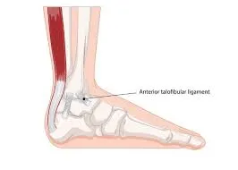
The ATFL is more than just a simple connective tissue; it's a sophisticated biological bridge that links two critical bones in the ankle complex. Anatomically positioned on the lateral aspect of the ankle, this ligament connects the fibula (the smaller of the two lower leg bones) to the talus (a key bone in the foot's articulation). Its precise location and unique structural composition make it particularly vulnerable to injury, yet simultaneously critical for maintaining ankle stability.
Detailed Ligament Characteristics:
| Characteristic | Specific Details |
|---|---|
| Length | 2-3 centimeters |
| Width | Approximately 10-12 millimeters |
| Thickness | Typically 1-2 millimeters |
| Primary Function | Ankle stabilization and movement restriction |
| Injury Vulnerability | Highest among ankle ligaments |
Biomechanical Role and Movement Dynamics
When we consider the complex movements of the human ankle, the ATFL emerges as a critical regulator of motion. It acts as a sophisticated biological restraint, preventing excessive inward rotation of the foot—a movement known in medical terms as inversion. This seemingly simple function is actually incredibly complex, involving intricate interactions between muscle, bone, and connective tissue.
During walking, running, or more dynamic activities like sports, the ATFL undergoes constant stress and strain. It must simultaneously provide stability and allow for the nuanced movements that make human locomotion so efficient and graceful. Imagine a finely tuned suspension system in a high-performance vehicle—this is essentially the role of the ATFL in our body's movement mechanism.
Common Injury Mechanisms and Risk Factors
Understanding ATFL injuries requires a holistic view of both mechanical and physiological factors. The ligament is most commonly injured during activities that involve sudden changes in direction, unexpected ground surfaces, or high-impact movements.
Risk Factors for ATFL Injury:
| Category | Specific Risks | Impact Level |
|---|---|---|
| Sports | Basketball, Soccer, Football | High |
| Recreational Activities | Hiking, Dancing | Medium |
| Occupational | Construction, Warehouse Work | Medium-High |
| Personal Factors | Previous Ankle Injuries | High |
| Physiological Considerations | Ankle Anatomy, Muscle Strength | Variable |
The Healing and Rehabilitation Journey
Recovery from an ATFL injury is not merely a medical process but a holistic journey of rehabilitation and understanding. The body's remarkable ability to heal is most evident in how ligaments can regenerate and strengthen with proper care and targeted interventions. Physical therapy becomes not just a treatment but a collaborative process between the patient's body and professional guidance.
A comprehensive rehabilitation approach typically involves multiple stages:
- Initial Protection and Rest
- Gradual Range of Motion Restoration
- Strength Rebuilding
- Functional Movement Retraining
- Progressive Return to Normal Activities
Psychological Aspects of Injury and Recovery
Beyond the physical dimensions of an ATFL injury lies a profound psychological landscape. Patients often experience anxiety, frustration, and uncertainty during recovery. Understanding that healing is a process—not an event—becomes crucial. The mental resilience required parallels the physical rehabilitation, making patient education and emotional support integral to successful recovery.
Medical Imaging and Diagnostic Visualization
Advanced Imaging Techniques for ATFL Assessment
Modern medical diagnostics have revolutionized our understanding of ligament injuries, with imaging technologies providing unprecedented insights into the intricate structures of the ankle. The anterior talofibular ligament (ATFL) requires specialized imaging approaches to fully comprehend its condition and potential injuries.
Imaging Modalities Comparison:
| Imaging Type | ATFL Visibility | Advantages | Limitations |
|---|---|---|---|
| X-Ray | Limited | Quick, Low Cost | Cannot directly visualize soft tissue |
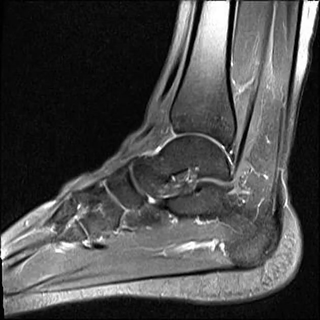
| MRI | Excellent | Detailed Soft Tissue Visualization | Expensive, Time-Consuming |
| Ultrasound | Good | Real-Time Imaging, Dynamic Assessment | Operator-Dependent |
| CT Scan | Moderate | Bone Structure Clarity | Radiation Exposure |

X-Ray Interpretation: A Critical Diagnostic Skill
X-ray interpretation for ATFL-related injuries requires a nuanced approach that goes beyond simple visual assessment. Professional radiologists and specialized tools like X-ray Interpreter follow a systematic methodology to detect subtle signs of ligament compromise. This careful analysis helps healthcare providers make accurate diagnoses and develop appropriate treatment plans.
Key X-Ray Interpretation Indicators:
- Talar Tilt Angle
- Joint Space Abnormalities
- Indirect Signs of Ligament Instability
- Comparative Analysis with Uninjured Side
Advanced X-Ray Interpretation Techniques:
- Digital Enhancement Protocols
- Multi-Angle Radiographic Assessment
- Comparative Measurement Techniques
Visual Diagnostic Image Gallery
To provide a comprehensive understanding, we've curated a specialized image collection demonstrating ATFL-related diagnostic visualizations:
Image Categories:
-
Normal ATFL Anatomy
- High-resolution MRI cross-sections
- 3D reconstructed ligament models
- Anatomical landmark illustrations
-
Injury Visualization
- Partial ligament tear representations
- Complete ligament rupture imaging
- Comparative healthy vs. injured ligament scans
-
Diagnostic Imaging Techniques
- X-ray stress view demonstrations
- Ultrasound dynamic assessment
- MRI soft tissue contrast images
Emerging Technological Innovations
The field of medical imaging continues to evolve, with cutting-edge technologies promising more precise ATFL diagnostic capabilities:
| Technology | Diagnostic Potential | Current Status |
|---|---|---|
| AI-Enhanced X-Ray Analysis | High Precision Detection | Emerging |
| 3D Volumetric Imaging | Comprehensive Visualization | Advanced Development |
| Molecular Imaging Techniques | Cellular-Level Assessment | Research Phase |
Case Studies: Real-World ATFL Injuries and Recovery
Case 1: The Weekend Athlete
Patient Profile:
- 32-year-old male
- Recreational basketball player
- Grade 2 ATFL sprain from landing awkwardly
Treatment Journey:
- Initial RICE protocol
- 6 weeks of physical therapy
- Gradual return to sports over 3 months
- Full recovery with preventive ankle exercises
Key Takeaway: Early intervention and consistent rehabilitation led to complete recovery without surgical intervention.
Case 2: The Ballet Dancer
Patient Profile:
- 24-year-old female
- Professional dancer
- Chronic ATFL instability
Treatment Journey:
- Conservative treatment initially
- Custom orthotics
- Modified dance technique
- Specialized strengthening program
- Return to performance in 4 months
Key Takeaway: Profession-specific rehabilitation approaches can be crucial for optimal recovery.
Case 3: The Senior Walker
Patient Profile:
- 68-year-old female
- Grade 1 sprain from uneven surface
- Pre-existing arthritis
Treatment Journey:
- Gentle mobility exercises
- Balance training
- Walking aid temporarily
- Gradual strength building
- Full recovery in 6 weeks
Key Takeaway: Age-appropriate treatment plans and patience are essential for successful outcomes.
Common Success Factors
Across these cases, several elements contributed to successful recovery:
- Early and accurate diagnosis
- Commitment to rehabilitation
- Patience with the healing process
- Professional guidance
- Modified activity during recovery
- Preventive measures post-recovery
Long-Term Management and Prevention Strategies
Preventing ATFL injuries transcends simple exercise routines. It encompasses a comprehensive approach to body awareness, biomechanical understanding, and proactive health management. This might involve:
- Regular flexibility and strength training
- Proper footwear selection
- Understanding individual biomechanical limitations
- Gradual progression in physical activities
- Consistent body awareness and movement mindfulness
Frequently Asked Questions
How long does it take for an anterior talofibular ligament to heal?
Healing time varies depending on the severity of the injury:
- Grade 1 (mild sprain): 2-4 weeks
- Grade 2 (moderate tear): 4-8 weeks
- Grade 3 (complete tear): 8-12 weeks Recovery time can be longer if proper rehabilitation protocols aren't followed.
Can an ATFL tear heal on its own?
Minor to moderate ATFL tears (Grade 1 and 2) can typically heal on their own with proper care, including rest, appropriate rehabilitation exercises, and following RICE protocol (Rest, Ice, Compression, Elevation). However, professional medical guidance is essential for proper healing.
Does a grade 3 ATFL tear need surgery?
Not always. While Grade 3 tears (complete ruptures) sometimes require surgical intervention, the decision depends on various factors:
- Patient's age and activity level
- Overall ankle stability
- Response to conservative treatment
- Professional/athletic requirements Many Grade 3 tears can be treated successfully with conservative management and proper rehabilitation.
Why is the ATFL the weakest ligament?
The ATFL is considered vulnerable due to several factors:
- Its anatomical position
- Thin structural composition
- Frequent stress during normal movement
- Limited blood supply These characteristics make it the most commonly injured ankle ligament.
How to heal ankle ligaments faster?
While you can't rush natural healing, you can optimize the process by:
- Following medical advice strictly
- Adhering to the RICE protocol
- Performing prescribed exercises
- Maintaining proper nutrition
- Getting adequate rest
- Avoiding re-injury during healing
Do ankle ligaments ever fully heal?
Yes, ankle ligaments can heal completely with proper care. However, the healed ligament may have slightly different characteristics than the original tissue. With appropriate rehabilitation, most people regain full functionality, though some may need to maintain strengthening exercises long-term.
Is walking good for torn ligaments?
Walking with a torn ligament depends on:
- The severity of the injury
- Stage of healing
- Medical professional's advice
Early after injury, rest is crucial. As healing progresses, gradual weight-bearing activities are typically introduced under professional guidance. Never force walking if it causes pain or instability.
Conclusion: Respecting the Complexity of Our Ankle
The anterior talofibular ligament represents far more than a mere anatomical footnote. It embodies the intricate, beautiful complexity of human movement—a testament to the sophisticated biological engineering that allows us to navigate the world with grace, stability, and resilience.
By understanding, respecting, and caring for this small yet significant ligament, we honor the remarkable machinery of our own bodies. The continuous advancement in medical understanding, diagnostic technologies, and treatment approaches gives us ever-improving tools to maintain and restore ankle health, ensuring we can stay active and mobile throughout our lives.