
How to Identify Errors and Inconsistencies in AI-Generated Diagnostic Reports
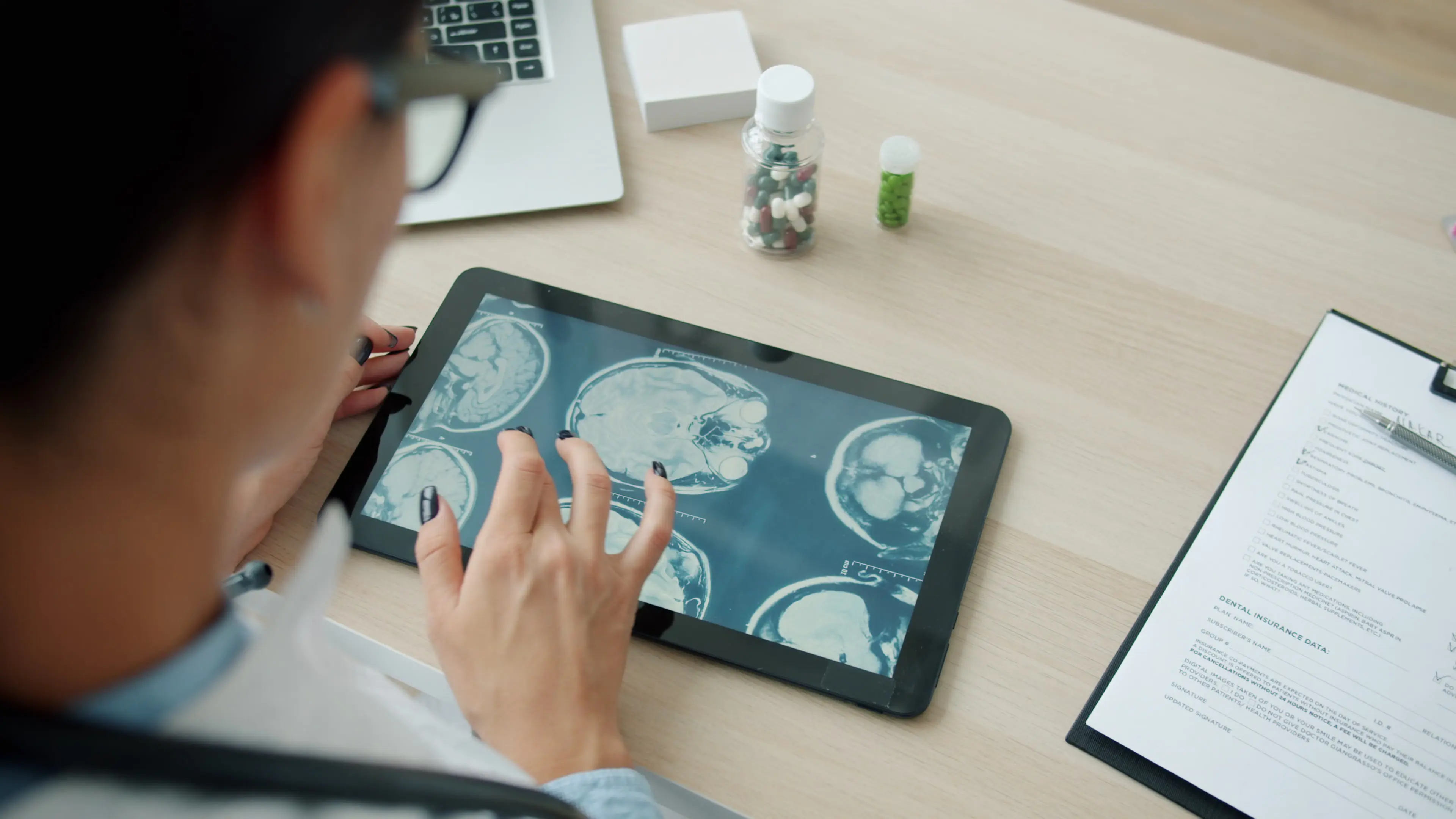
For years, radiologists and technologists have spent hours reading CT slices and MRIs to write reports.
Lately, AI has been employed to help write diagnostic reports, promising faster drafts and less work. This technology is useful until the polished text quietly invents a finding, swaps left for right, or misses something subtle.
As Charles Kahn, editor of Radiology: Artificial Intelligence, aptly noted regarding these modern dilemmas, “Is that really an abnormality, or am I missing something that AI, with its subtle mind or whatever, is identifying? And that is really challenging for us.”
This is why knowing how to spot errors and inconsistencies in AI-generated reports has become essential for imaging specialists and healthcare professionals who rely on them.
In this article, we will discuss the most common diagnostic errors and tips to help you spot errors and inconsistencies in AI-generated reports.
Common Types of AI Diagnostic Errors
AI doesn’t see images the way you do after years of training. It predicts patterns from data, which leads to predictable problems.
A 2025 systematic review on ScienceDirect examined 15 studies of large language models writing radiology reports. It found the same issues over and over. Hallucinations (made-up findings) showed up in 40% of the studies, mis- or over-diagnosis in 53%, and missing clinical details in 80%.
Here are some common types of AI diagnostic errors that occur repeatedly:
-
Hallucinations (anatomical, pathological, and measurement): These include invented anatomical structures, phantom lesions that don’t exist on the actual images, or wrong size measurements (for example, calling a lesion 3.2 cm when it is actually 4.1 cm).
-
Laterality, location, and anatomical mislabeling: Models frequently swap left and right, misidentify structures, or place findings in the wrong compartment. These errors appear in both pure language models and vision-language systems.
-
Inconsistencies between findings and impressions: The report can sound fluent while the impression quietly contradicts the described findings or invents management advice that doesn’t match the images.
-
Missed findings and false negatives: Subtle or uncommon abnormalities often go undetected, especially in cases where the model simply fails to flag something a trained eye would catch.
How to Identify Errors and Inconsistencies in AI-Generated Diagnostic Reports
Here are three areas you must focus on to identify errors and inconsistencies in AI-generated reports:
Verify Biomarker Ranges Against Current Guidelines
AI may report a lesion diameter, an ejection fraction estimate, or a laboratory biomarker range that appears precise. What you must do is treat every number as provisional until you verify it against the image and current specialty guidelines.
Pull the actual measurement tools or prior reports. Compare stated sizes against Fleischner, Breast Imaging Reporting and Data System (BI-RADS), or whatever applies for the organ. Check whether a “mild” or “severe” label matches published thresholds.
If the report cites a range or cutoff, confirm it against the latest society guideline rather than accepting the model’s memory. Models trained on older data can quote outdated thresholds. Cross-check any invented serial measurements against the actual prior study.
Audit Against Population and Historical Baselines
Even when the numbers look guideline-compliant, they can still be inconsistent with reality.
Models trained on narrow datasets may produce findings whose prevalence or severity does not reflect the local population or whose details conflict with the patient's previous imaging.
Compare the current report to the patient’s own prior imaging and reports. Sudden appearance of a finding, or disappearance of a documented abnormality without treatment, needs explanation.
Note demographic mismatches as well. Pediatric, non-Western, or multilingual populations are underrepresented in many public datasets.
Family nurse practitioners (FNPs) can be of great help here. American International College notes that FNPs handle physical exams, order and interpret tests, prescribe medications, and build treatment plans.
These professionals typically hold a graduate degree in nursing with an FNP specialization. They bring a longitudinal primary-care perspective on symptoms, labs, and patient history that can be cross-checked against the imaging summary.
Even graduates who pursue an online master’s FNP degree program to maintain clinical hours or jobs while studying also develop sharp diagnostic instincts. It’s because of the rigorous clinical training that they can immediately spot when a health pattern drifts from expected community norms or an individual patient’s history.
Look for Phantom Artifacts in Imaging Summaries
Phantom findings, such as lesions or nodules that exist only in the AI text, are among the most dangerous because they sound entirely convincing. Research published in PMC identifies phantom lesions as accounting for 8 - 15% of hallucination examples.
Cross-check every positive statement against the actual images. If the report mentions a “left apical nodule” or “right adrenal adenoma” that you cannot locate, mark it. Pay special attention to laterality swaps and to descriptions of enhancement or signal characteristics that do not match the sequences provided.
Be careful with synthetic or heavily post-processed images. The 2026 RSNA Margulis Award study showed that even experienced radiologists identified AI-generated X-rays only about 70–75% of the time after being told AI-generated ones were present.
Diagnostic accuracy on the fakes was similar to real images, meaning the fakes can fool both eyes and models.
FAQs
What are AI hallucination errors in radiology reports?
AI hallucinations occur when systems generate fabricated anatomical details, incorrect measurements, or non-existent lesions in radiology reports. These descriptions do not match the actual scans. Such errors can mislead clinicians, potentially resulting in incorrect diagnoses, unnecessary tests, or inappropriate treatment decisions.
How do prior medical histories help spot AI reporting errors?
Comparing AI-generated summaries with previous imaging reports helps identify inconsistencies that require further investigation. For example, a large lesion might suddenly appear, or a chronic abnormality might disappear without treatment. Reviewing patient histories helps radiologists recognize these discrepancies and verify findings before finalizing reports.
Why is clinical oversight necessary for AI diagnostic drafts?
AI models can misinterpret complex imaging patterns, overlook subtle abnormalities, or reference outdated clinical thresholds. Qualified radiologists must review AI-generated drafts before approving reports. Their expertise helps verify findings, correct inaccuracies, and ensure diagnostic conclusions support appropriate patient care and treatment decisions.
Key Statistics
| Description / Finding | Statistic |
|---|---|
| Prevalence of AI hallucinations (made-up findings) in radiology report studies | 40% |
| Prevalence of mis- or over-diagnosis in reviewed AI studies | 53% |
| Prevalence of missing clinical details in AI-generated reports | 80% |
| Frequency range of phantom lesions within AI hallucination examples | 8% to 15% |
| Rate at which experienced radiologists successfully identified AI-generated X-rays after being told they were present | 70% to 75% |
You do not have to catch every possible error on the first pass. Focus first on numbers that sit near decision thresholds, on patterns that feel off for the patient or population, and on any positive finding you cannot see. Those three habits alone catch most of the problems that matter.
Treat every AI draft as a helpful first pass, then apply the clinical judgment you have built over years of reading images and knowing patients. The technology will keep improving, but your careful review is what keeps reports safe and useful.