
Beyond the X-ray: How Different Imaging Technologies Support Modern Diagnosis
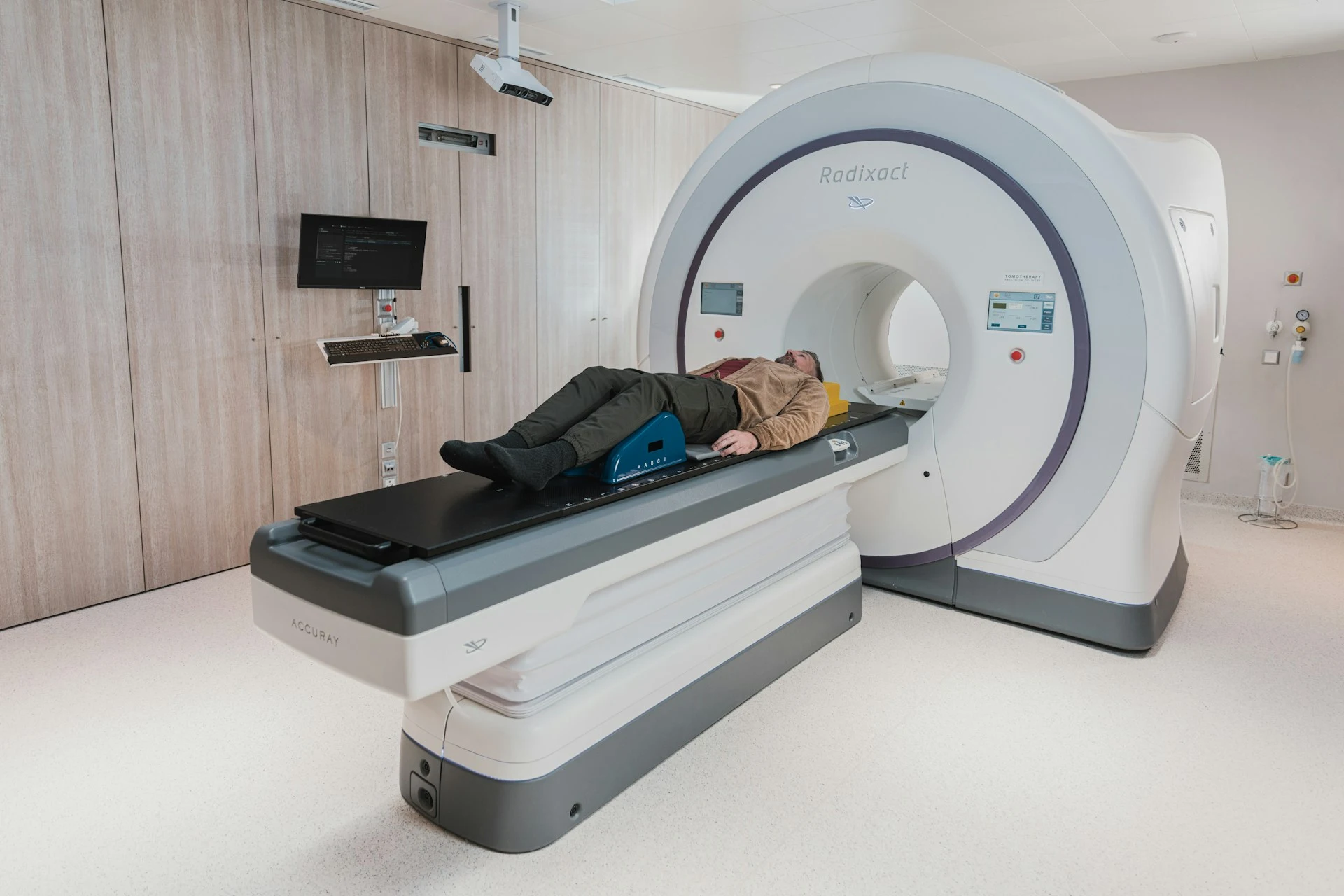
If you’ve ever broken a bone, injured your ankle, or complained of persistent pain, there’s a good chance a healthcare professional has sent you for a scan. It’s one of the most familiar diagnostic tests, and for good reason. It gives doctors a quick look at what’s happening inside the body without requiring surgery or other invasive procedures.
And the scale of medical imaging is pretty remarkable. Industry reports suggest that more than 607 million imaging procedures are carried out every year in the U.S. alone. But X-rays account for only part of that picture. The number also includes other imaging methods you may not be as familiar with.
But why are there so many different imaging methods, and what makes each one different? Let’s take a closer look.
Why One Imaging Method Isn’t Enough
The short answer is that no single scan shows everything.
A plain X-ray does a good job with bones and some chest problems. But if your joint pain won’t quit, or someone suspects a torn ligament, an MRI is often the better call. When you’re pregnant, your doctor may order an ultrasound to see how your baby is growing and catch any health issues early. Different problems call for different imaging procedures.
So who actually reads these images? Radiologists do, mostly, along with other physicians and dentists for dental scans. Your typical registered nurse doesn’t. However, advanced practice nurses, like nurse practitioners, can. The American Association of Nurse Practitioners, for example, says that nurse practitioners can assess patients, order and interpret diagnostic tests, make diagnoses, and manage treatment plans. This is, of course, subject to state regulations.
Nurses who want to move into this type of advanced practice have options. A 100% online FNP program is one possible route into family nurse practitioner work. What’s more, many of these programs can be completed in a relatively short time.
Rockhurst University, for instance, says its full-time FNP program runs six semesters, and the part-time option runs eight.
Still, finishing an FNP program doesn’t automatically mean someone can sign off on formal radiology reports. But it does move them closer.
Types of Medical Imaging Technologies and What They Reveal
Let’s walk through the most common imaging procedures and what each can show.
X-rays: Examining Bones and Chest Abnormalities
X-rays are commonly used to identify fractures, dislocations, dental cavities, and chest conditions such as pneumonia.
For example, a chest X-ray can help doctors find signs of pneumonia or other lung problems. Dentists also take X-rays of your teeth to spot cavities, infections, and problems with the roots of teeth that can’t be seen during a routine examination. In fact, dental X-rays made up about 320 million radiographic exams in 2016, according to the JAMA Network.
Their main limitation is soft tissue detail. While X-rays can reveal some abnormalities beyond the skeleton, they’re not always enough to investigate more complicated conditions.
CT Scans: Taking a Closer Look at Internal Structures
A computed tomography scan, or CT scan for short, is essentially an advanced version of the X-ray. It takes X-ray images from various angles and assembles them into layered cross-sections. This helps in identifying internal bleeding, traumatic injuries, blood clots in the lungs, as well as certain cancers.
CT scans are also very important during emergencies when time is of the essence. ”In the ER, time isn't just money. It’s neurons,” wrote radiologist Dr. Muhammad Naeem on LinkedIn on why head CT scans are the ”MVP” of the ER. ”When a patient rolls in with an altered mental status, a sudden ”worst headache of their life,” or focal weakness, the non-contrast CT head is our first line of defense.”
The downside is the risk of cancer. JAMA Network estimated that the 93 million CT scans performed in 2023 could likely result in 103,000 future cancers. These are projected cases, not confirmed diagnoses.
MRI: Seeing Soft Tissues and Complex Structures
MRI, or magnetic resonance imaging, uses powerful magnets and radio waves to map out the inside of your body. The images can be of the brain, spinal cord, muscles, ligaments, and joints. They come out really sharp so doctors can see exactly what’s going on.
Say you’ve got a suspected torn ACL or a slipped disc in your spine. An MRI gives your doctor a far better view, making it far more effective when doctors want to see what standard X-rays can barely show.
There are more than 100 million MRI scans each year worldwide, reflecting just how important the technology has become in modern diagnosis.
Ultrasound: Seeing What’s Happening Inside the Body in Real Time
Ultrasound, called medical ultrasonography in official circles, uses high-frequency sound waves to build live images of organs and blood flow on a screen.
Ultrasound is popular as a pregnancy scan procedure, but it goes well beyond that. Doctors use it to check abdominal organs and thyroid nodules, as well as assess vascular health through Doppler techniques. Its biggest upside is that it doesn’t use ionizing radiation, yet it still delivers images in real time.
The numbers show how heavily it’s relied on. A 2024 study in the Journal of Ultrasound in Medicine found that outpatient ultrasound exams in the US climbed from 38.6 million in 2011 to 59.8 million in 2021.
Quick Comparison: What Do Different Imaging Scans Show?
| Imaging Technology | What It Reveals | Common Uses |
|---|---|---|
| X-ray | Bones & some chest abnormalities | Fractures, dental problems, pneumonia |
| CT Scan | Clear, cross-sectional images of what is inside the body | Internal bleeding, injuries, blood clots |
| MRI Scan | Soft tissues, organs, brain, & spinal structures | Ligament injuries, spinal conditions, brain abnormalities |
| Ultrasound Scan | Organs, soft tissues, & blood flow | Pregnancy monitoring, abdominal examinations, vascular problems |
FAQs
Which imaging test is the safest?
Ultrasound and MRI don’t use ionizing radiation, so they’re typically safe. Radiation and the risk of cancer are the main concerns with X-rays and CT scans. That said, ”safest” depends on what your doctor needs to find.
Why would my doctor choose a CT scan over an X-ray?
A doctor will usually pick a CT scan over a standard X-ray when they require a far more detailed three-dimensional cross-sectional view of what’s wrong inside the body. X-ray works well, but it’s limited in what it can reveal to doctors.
Can a nurse read my scan?
Yes, but it depends on the type of nurse. Diagnostic scans are usually read and interpreted by a radiologist or the doctor who ordered the scan. However, in many states, as well as in some urgent care settings, trained nurse practitioners may review X-rays for preliminary findings.
Different Technologies, One Diagnostic Goal
X-rays, CT scans, MRIs, and ultrasounds each offer a different lens on the human body. Each scan procedure has its strengths as well as its limitations, as we’ve seen in this guide. But used in the right scenario, they give healthcare professionals a range of ways to investigate what’s going on inside you.
The real value isn’t just in the machine. It’s in the judgment of the person who orders the right test and the expert who reads it.