A label-free optical imaging technique using autofluorescence lifetime and AI can distinguish colorectal cancer with 85% accuracy.
Key Details
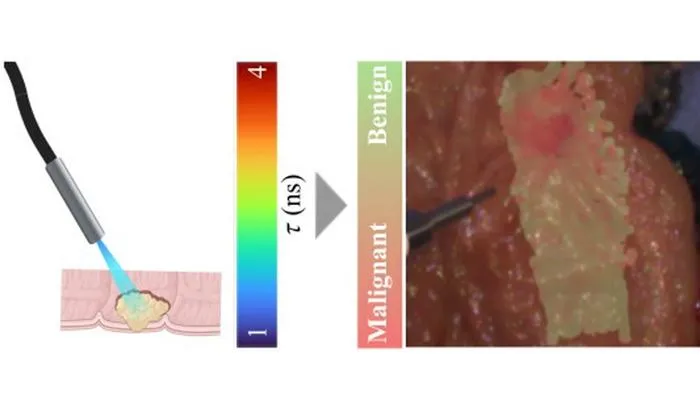
- 1Champalimaud Foundation researchers developed a fiber-optic, label-free optical imaging method for colorectal tissue analysis.
- 2Technique involves autofluorescence lifetime measurements at two wavelengths to capture biochemical differences.
- 3Machine learning (AdaBoost) trained on 117 patients' surgical specimens, validated with matched pathology results.
- 4On test data, the AI achieved 85% accuracy, 85% sensitivity, and 85% specificity.
- 5Potential applications include real-time cancer detection during colonoscopy or surgery, reducing the need for biopsies.
- 6Simplified versions of the imaging system delivered strong results, supporting future clinical use.
Why It Matters
Real-time, label-free optical imaging enhanced with AI could support faster, more accurate detection of cancer during endoscopic procedures, leading to earlier intervention and fewer unnecessary biopsies. This advances integration of functional imaging and AI in clinical workflows.

Source
EurekAlert
Related News

•EurekAlert
AI Accelerates Radiopharmaceuticals, Boosts Personalized Dosimetry in Cancer
Machine learning is driving advancements in radiopharmaceutical drug discovery and optimizing patient-specific dosimetry for precision cancer therapy.

•EurekAlert
Physicians Overly Trust Erroneous AI, Ignore Contradictory Evidence
Physicians tend to trust incorrect AI advice, even when evidence contradicts it, suggesting risks in clinical decision-making with AI tools.

•EurekAlert
Concerns Raised Over Unverified Datasets in AI Health Prediction Models
A new study finds widely used AI health prediction models are built on datasets with unverifiable origins, raising safety and validity concerns.