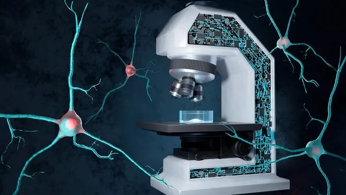
EPFL researchers created an AI-driven microscopy system that predicts and analyzes misfolded protein aggregation in real time.
Key Details
- 1Developed a self-driving imaging system combining multiple microscopy methods and deep learning.
- 2System predicts and detects protein aggregation—a hallmark of neurodegenerative diseases—in living cells.
- 3Uses label-free microscopy to minimize sample alteration and maximize imaging efficiency.
- 4Upon aggregation detection, system triggers Brillouin microscope to analyze biomechanical properties of aggregates.
- 5Aggregation onset detection achieved 91% accuracy using a specialized deep learning algorithm.
- 6Published in Nature Communications, with potential impact on drug discovery and precision medicine.
Why It Matters

Source
EurekAlert
Related News

Deep Learning AI Outperforms Clinic Prognostics for Colorectal Cancer Recurrence
A new deep learning model using histopathology images identifies recurrence risk in stage II colorectal cancer more effectively than standard clinical predictors.

AI Reveals Key Health System Levers for Cancer Outcomes Globally
AI-based analysis identifies the most impactful policy and resource factors for improving cancer survival across 185 countries.

Deep Learning Boosts ICD-11 Coding Accuracy for Chinese EMRs
Researchers developed a deep learning model achieving high accuracy in automatic ICD-11 coding of Chinese electronic medical records.