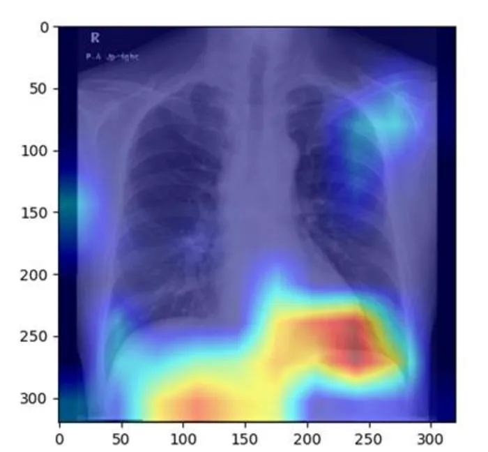
Researchers developed an AI model that can accurately detect fatty liver disease from routine chest X-rays.
Key Details
- 1The AI model was created by Osaka Metropolitan University researchers.
- 2It was trained and validated with 6,599 chest X-ray images from 4,414 patients.
- 3Model performance was strong, with AUC between 0.82 and 0.83.
- 4Chest X-rays are less costly and more commonly performed than ultrasounds, CTs, or MRIs currently used for liver diagnosis.
- 5Results were published in Radiology Cardiothoracic Imaging on June 20, 2025.
Why It Matters
This approach could enable earlier and more widespread detection of fatty liver disease using existing chest X-rays, reducing the need for more expensive or specialized imaging modalities. It demonstrates the potential for AI to repurpose common imaging studies for new clinical insights in radiology.

Source
EurekAlert
Related News

•EurekAlert
AI Accelerates Radiopharmaceuticals, Boosts Personalized Dosimetry in Cancer
Machine learning is driving advancements in radiopharmaceutical drug discovery and optimizing patient-specific dosimetry for precision cancer therapy.

•EurekAlert
Physicians Overly Trust Erroneous AI, Ignore Contradictory Evidence
Physicians tend to trust incorrect AI advice, even when evidence contradicts it, suggesting risks in clinical decision-making with AI tools.

•EurekAlert
Concerns Raised Over Unverified Datasets in AI Health Prediction Models
A new study finds widely used AI health prediction models are built on datasets with unverifiable origins, raising safety and validity concerns.