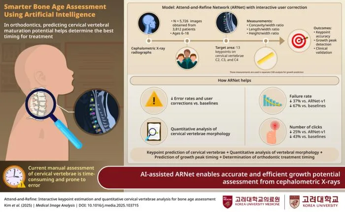
Korean researchers developed an AI system (ARNet-v2) that predicts children's growth spurts from neck X-rays to enhance orthodontic treatment planning.
Key Details
- 1ARNet-v2 uses lateral cephalometric radiographs to identify cervical vertebrae keypoints.
- 2The model allows a single clinician correction to propagate, boosting efficiency and accuracy.
- 3Tested on 5,700+ radiographs and across four public datasets, ARNet-v2 reduced prediction failures by up to 67%.
- 4Manual annotation requirements are halved compared to conventional approaches.
- 5The AI may reduce the need for additional hand–wrist X-rays, lowering radiation exposure for pediatric patients.
- 6Published in Medical Image Analysis, July 2025.
Why It Matters
This model offers significant efficiency and diagnostic accuracy improvements for pediatric orthodontics, potentially lowering cost and radiation exposure for young patients. Its success could pave the way for further AI integration in radiology workflows and broader medical imaging challenges.

Source
EurekAlert
Related News

•EurekAlert
AI Accelerates Radiopharmaceuticals, Boosts Personalized Dosimetry in Cancer
Machine learning is driving advancements in radiopharmaceutical drug discovery and optimizing patient-specific dosimetry for precision cancer therapy.

•EurekAlert
Physicians Overly Trust Erroneous AI, Ignore Contradictory Evidence
Physicians tend to trust incorrect AI advice, even when evidence contradicts it, suggesting risks in clinical decision-making with AI tools.

•EurekAlert
Concerns Raised Over Unverified Datasets in AI Health Prediction Models
A new study finds widely used AI health prediction models are built on datasets with unverifiable origins, raising safety and validity concerns.