A new AI tool estimates bone mineral density from routine lumbar spine and femur X-rays, enabling earlier osteoporosis detection.
Key Details
- 1Traditional osteoporosis screening uses dual-energy X-ray absorptiometry (DXA) but has low utilization rates.
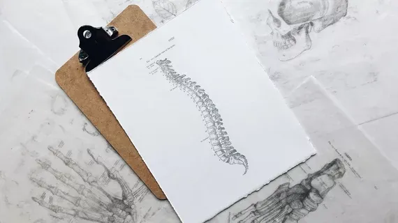
- 2The AI estimates bone mineral density from lumbar spine and femur X-rays already performed for other indications.
- 3Experts believe this may help identify at-risk patients sooner for earlier intervention.
- 4Millions are currently undiagnosed due to limited access to DXA equipment.
- 5Details of the development were published in the Journal of Orthopaedic Research.
Why It Matters
By leveraging commonly performed X-rays, this AI tool could expand osteoporosis screening to a broader population, addressing a major underdiagnosis issue in bone health and improving patient outcomes globally.

Source
Health Imaging
Related News

•Radiology Business
Framework Assesses Real-World Financial Impact of Radiology AI Adoption
A new analysis presents a financial calculator for objectively assessing the return on investment (ROI) of implementing radiology AI solutions.

•Radiology Business
AI Technique Unveils Previously Hidden MS Gray Matter Lesions on MRI
Researchers developed an AI-enhanced method to detect previously invisible gray matter lesions in multiple sclerosis using MRI.

•Radiology Business
Majority of Patients Want Disclosure When AI Used in Imaging
A new survey finds that nearly all patients want to be informed when AI is utilized in medical imaging interpretation.