Researchers have developed an AI-enhanced three-phase CT perfusion protocol that reduces radiation exposure by over 80% while accurately generating perfusion maps for stroke evaluation.
Key Details
- 1Traditional CT perfusion (CTP) uses continuous scanning, resulting in high radiation doses (~5260 mGy·cm) and workflow complexity.
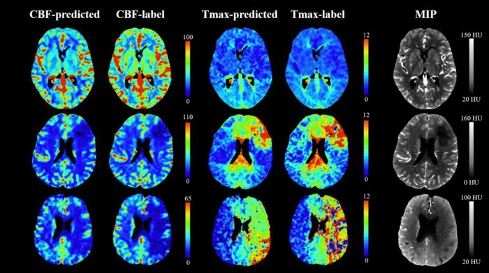
- 2The new protocol samples only three phases and uses a GAN-based deep learning model to generate cerebral blood flow (CBF) and Tmax maps.
- 3The approach reduces patient radiation exposure by more than 80% compared to standard CTP methods.
- 4Internal validation demonstrated high fidelity of AI-generated blood flow maps, even with slight deviations in timing of image acquisition.
- 5The method preserves diagnostic accuracy essential for stroke management and is more robust to patient motion.
Why It Matters
The innovation could make stroke diagnosis safer and more accessible, significantly lowering radiation risks while maintaining diagnostic efficacy. Adopting this protocol may benefit vulnerable populations, improve workflow, and expand access to functional brain imaging in acute care.

Source
EurekAlert
Related News

•EurekAlert
AI Accelerates Radiopharmaceuticals, Boosts Personalized Dosimetry in Cancer
Machine learning is driving advancements in radiopharmaceutical drug discovery and optimizing patient-specific dosimetry for precision cancer therapy.

•EurekAlert
Physicians Overly Trust Erroneous AI, Ignore Contradictory Evidence
Physicians tend to trust incorrect AI advice, even when evidence contradicts it, suggesting risks in clinical decision-making with AI tools.

•EurekAlert
Concerns Raised Over Unverified Datasets in AI Health Prediction Models
A new study finds widely used AI health prediction models are built on datasets with unverifiable origins, raising safety and validity concerns.