University of Arizona researchers achieved nearly 90% accuracy in pancreatic cancer phenotyping using label-free optical microscopy with deep learning AI.
Key Details
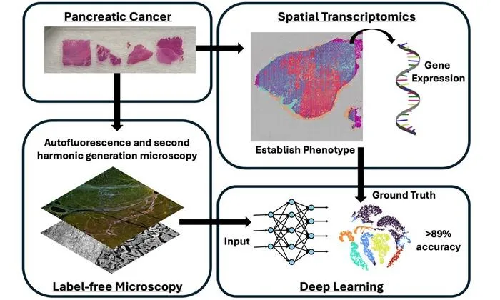
- 1Label-free optical microscopy paired with deep neural networks identified tissue phenotypes at over 89% accuracy in pancreatic cancer samples.
- 2Spatial transcriptomics served as the 'ground truth' for phenotypic classification.
- 3Traditional image analysis could not match the performance of AI methods, pointing to AI's necessity in extracting meaningful features from label-free images.
- 4This approach bypasses expensive and time-intensive molecular/genetic sequencing currently used in precision medicine.
- 5The work demonstrates a significant step toward more accessible and rapid phenotyping for cancer care.
Why It Matters
This study advances the use of imaging AI in precision medicine by offering a faster, less costly method for cancer phenotyping. It highlights the potential for optical imaging and AI to broaden access to personalized care by reducing dependence on resource-heavy molecular diagnostics.

Source
EurekAlert
Related News

•EurekAlert
AI Accelerates Radiopharmaceuticals, Boosts Personalized Dosimetry in Cancer
Machine learning is driving advancements in radiopharmaceutical drug discovery and optimizing patient-specific dosimetry for precision cancer therapy.

•EurekAlert
Physicians Overly Trust Erroneous AI, Ignore Contradictory Evidence
Physicians tend to trust incorrect AI advice, even when evidence contradicts it, suggesting risks in clinical decision-making with AI tools.

•EurekAlert
Concerns Raised Over Unverified Datasets in AI Health Prediction Models
A new study finds widely used AI health prediction models are built on datasets with unverifiable origins, raising safety and validity concerns.