如何解讀關節超聲波:3 種基本方法
December 13, 2024
關節超聲波是一種重要的影像技術,用於評估關節及其周圍組織。本指南將探討關節超聲波能顯示什麼、為什麼使用它,以及你可以如何解讀結果。
什麼是關節超聲波?
關節超聲波是一種非侵入性的影像方法,利用聲波即時生成關節、韌帶、肌腱和軟組織的影像。它安全、無痛,且不使用輻射。讓我們了解這種技術的內容以及它能顯示什麼。
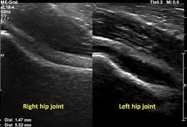
關節超聲波能顯示什麼?
- 關節積液: 檢測關節腔內異常的液體積聚。
- 肌腱問題: 可視化肌腱炎、撕裂或關節周圍肌腱的增厚。
- 韌帶損傷: 幫助識別扭傷、撕裂和韌帶的炎症。
- 軟骨評估: 提供對軟骨完整性的有限評估。
- 軟組織異常: 確認軟組織中的腫塊、囊腫或炎症。
- 骨表面不規則: 可以顯示表面變化和骨頭外生。
- 即時評估: 允許在關節運動時進行動態成像。
為什麼你可能需要關節超聲波?
- 關節疼痛: 評估不明原因的關節不適的原因。
- 腫脹: 評估關節積液和軟組織炎症。
- 活動範圍受限: 幫助識別運動限制的來源。
- 運動傷害: 診斷與肌腱、韌帶和肌肉相關的傷害。
- 後續監測: 檢查已知關節疾病的進展。
- 引導程序: 為關節注射或抽取引導針頭位置。
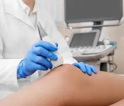
如何進行關節超聲波檢查?
關節超聲波是一個快速且無痛的程序。以下是你可以期待的內容:
- 準備: 你可能需要脱去任何珠寶或覆蓋要檢查區域的衣物。
- 擺位: 你會舒適地擺放,根據被掃描的關節坐著或躺下。
- 施加凝膠: 將透明的水性凝膠塗抹在關節上方的皮膚上。這種凝膠有助於聲波有效傳播。
- 掃描: 一個看起來像小型麥克風的探頭將在塗有凝膠的區域上移動。這個探頭發出聲波並接收回聲,實時在顯示器上形成影像。
- 影像回顧: 超聲檢查師或醫生將檢視影像,評估關節及其周圍組織是否有異常。
- 清理: 凝膠會被擦掉,你可以在掃描後立即回到你的活動中。
如何解讀關節超聲波結果
解讀關節超聲波需要理解正常解剖結構以及識別病理變化。以下是三種解讀的方法。
1. 使用 X-ray Interpreter
X-ray Interpreter 提供 AI 驅動的分析來協助進行超聲解讀:
- 註冊: 在 X-ray Interpreter 上註冊以使用我們的 AI 進行超聲分析。
- 上傳影像: 將你的關節超聲影像上傳到平台。
- 查看解讀: 接收 AI 生成的解讀報告。
- 諮詢: 始終與你的醫生討論結果。
查看我們的 入門指南 獲取更多詳情。
2. 使用 ChatGPT Plus
ChatGPT Plus 也可以協助分析超聲影像,搭配它的 GPT-4V 模型:
- 訂閱: 訂閱 ChatGPT Plus 以獲取高級分析。
- 上傳影像: 將你的超聲影像上傳到 OpenAI 平台。
- 請求分析: 請求 AI 解讀超聲影像。
- 檢視和驗證: 檢視結果,始終與醫療專業人員確認。
在我們的 部落格 中了解如何使用 ChatGPT Plus 進行醫學影像解讀。
此外,隨著幾種其他擁有視覺能力的 AI 模型的出現,你也可以嘗試其他模型,如 Grok 由 xAI, Claude 由 Anthropic, Gemini 由 Google Deepmind。
3. 自行理解基礎知識
雖然不能取代醫療專業人士,但一些基本理解可以幫助你更好地理解你的結果。
- 學習解剖學: 熟悉基礎的關節解剖結構。
- 查看簡單指南: 使用在線資源了解關節超聲的常見發現。
- 記下問題: 記下不熟悉的術語並向你的醫療提供者詢問。
- 尋求專家指導: 始終與醫療專業人士確認你的理解。
比較不同的解讀方法
以下是解讀關節超聲波的不同方法的比較:
| 標準 | X-ray Interpreter | ChatGPT Plus | 自我閱讀 |
|---|---|---|---|
| 準確性 | 高 (基於 AI)1 | 高 (基於 AI)1 | 變化 (依賴技能) |
| 使用難易度 | 簡單 | 中等 | 具挑戰性 |
| 成本 | 每張影像起價 $2.50 | 每月 $20 | 免費 (不包括教育費用) |
| 時間效率 | 快速 | 中等到快速 | 慢到中等 |
| 學習曲線 | 低 | 低到中等 | 高 |
| 附加資源 | 提供 | 部分提供 (通過 OpenAI) | 自行獲取 |
每種方法都有其優勢和劣勢。AI 選項快速且精確,而基本的理解則能促進患者與醫生之間的更好溝通。
結論
關節超聲波是診斷各種肌肉骨骼疾病的有價值工具。本指南介紹了關節超聲技術的使用、它們生成的影像以及你可以更好理解結果的方法。
在選擇方法時,考慮你的需求、理解程度和可用資源。始終注意隱私標準,並尋求專業的醫學驗證。