目の超音波を解釈する方法: 3つの主要な方法
目の超音波、または眼球超音波は、音波を使用して目および周囲の組織の画像を生成する非侵襲的な画像診断方法です。このガイドでは、目の超音波が何を明らかにできるか、なぜ使用されるのか、そしてその結果をどのように理解するかを説明します。
目の超音波とは?
目の超音波は、目の内部の構造をリアルタイムで画像化する画像検査です。音波を利用し、安全で痛みのない手続きです。この技術がどのようなもので、何を示すことができるのかを探ってみましょう。
目の超音波で何がわかるか?
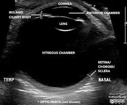
- 詳細な目の構造画像: 網膜、レンズ、視神経、硝子体の明確な可視化。
- 目の異常の検出: 網膜剥離、腫瘍、異物、その他の問題を特定する。
- 血流の評価: ドプラー超音波を使用して、目の血管の血流を評価することができる。
- リアルタイムイメージング: 目の構造や動きの動的な観察を可能にする。
- 手技のガイダンス: 注射やその他の治療の指針として利用可能。
なぜ目の超音波が必要なのか?
- 視力の問題: 視力の変化や問題があり、目の検査だけでは完全には診断できない場合。
- 目の痛みや怪我: 目の外傷やその他の痛みの原因を評価し診断するため。
- 疑わしい眼疾患: 緑内障や白内障のような状態を調査し、監視するために使用。
- 手術前の評価: 白内障手術などの手技の前に目の構造を評価するために使用。
- フォローアップの画像: 知られている目の状態の進行を監視するために。
目の超音波はどのように行われるか?
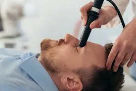
目の超音波は、通常15〜30分で完了する簡単で痛みのない手続きです。以下は、期待される一般的な流れです。
- 準備: メガネやコンタクトレンズを外すように求められます。テストのために座っているか、横になっているかのどちらかです。
- ジェルの塗布: まぶたに透明なジェルが塗布されます。このジェルは、超音波トランスデューサーが目と良好な接触を確保し、明確な画像を提供します。
- トランスデューサーの配置: トランスデューサーと呼ばれる小型の手持ち機器が、閉じたまぶたの上で優しく動かされます。トランスデューサーは、高周波の音波を発し、それを使用して目の画像を作成します。
- 画像取得: 超音波画像はリアルタイムでモニターに表示され、技術者または医師は眼球内および周囲の組織の構造を評価します。
- 完了: 必要な画像がキャプチャされると、ジェルが拭き取られ、手続きは完了です。
目の超音波結果の解釈方法
超音波結果を理解することは、目の健康を効果的に管理するための鍵です。以下は、結果の解釈を支援する一般的な方法の概要です。
1. X-ray Interpreterの利用
X-ray Interpreterは、AI駆動の分析を用いて超音波画像を分析します。以下の手順で使用できます。
- 登録: X-ray Interpreterにサインアップして、超音波分析のためのAIを利用します。
- 超音波のアップロード: 目の超音波画像をアップロードします。
- 解釈の確認: AIによって生成された解釈とレポートを受け取ります。
- 相談: 包括的な診断のために、必ず医師に相談してください。
詳細については、始め方ガイドをご覧ください。
2. ChatGPT Plusの利用
ChatGPT Plusは、その高度なGPT-4Vモデルを使用して超音波画像の分析を支援できます。
- サブスクリプション: 高度な分析のためにChatGPT Plusにサブスクライブします。
- 超音波のアップロード: OpenAIプラットフォームに超音波画像をアップロードします。
- 分析のリクエスト: AIに超音波画像を解釈し、レポートを提示するように依頼します。
- 確認と検証: 結果をレビューし、医療専門家にその正確性を確認します。
医療画像の解釈にChatGPT Plusを使用する方法については、ブログで詳細をご確認ください。
また、視覚能力を持つ他のAIモデルが数多く登場しているため、
xAIのGrokや、
AnthropicのClaude、
Google DeepmindのGeminiなど、他のモデルを試してみるのも良いでしょう。
3. 基本を自分で理解する
医療専門家の代わりにはなりませんが、基本を理解することで結果をより良く理解し、医師への質問を用意するのに役立ちます。
- 解剖学を学ぶ: 目の基本的な解剖学に慣れ親しんでください。
- 簡単なガイドを読む: 多くのオンラインリソースが、超音波の一般的な所見を理解するのに役立ちます。
- 質問する: 不明な用語をメモし、フォローアップ時に医療提供者に尋ねてください。
- 専門家のガイダンスを求める: 常に医療専門家に理解を確認してください。
様々なアプローチの比較
目の超音波の解釈に関するさまざまな方法を比較してみましょう。
| 基準 | X-ray Interpreter | ChatGPT Plus | 自己読解 |
|---|---|---|---|
| 精度 | 高 (AIベース)1 | 高 (AIベース)1 | 変動 (スキル依存) |
| 使いやすさ | 簡単 | 中程度 | 挑戦的 |
| 費用 | 画像ごとに$2.50から | 月額$20 | 無料 (教育費を除く) |
| 時間効率 | 速い | 中程度から速い | ゆっくりから中程度 |
| 学習曲線 | 低 | 低から中程度 | 高 |
| 追加リソース | 提供される | 部分的に提供 (OpenAIを通じて) | 自己調達 |
各方法には利点と欠点があります。AI駆動のオプションは速くて正確ですが、基本的な理解は患者と医師のコミュニケーションを助けます。
結論
目の超音波は、さまざまな医療目の状態を診断するための貴重なツールです。このガイドでは、超音波技術の使用、生成される画像、およびAIツールや自己学習を通じて結果をより良く理解する方法について紹介しました。
方法を選ぶ際には、特定のニーズや理解のレベル、利用可能なリソースを考慮してください。常にプライバシー基準を遵守し、専門的な医療確認を求めてください。