Wie man die Diffusionstensorbildgebung interpretiert: 3 wesentliche Techniken
Das Verständnis der Diffusionstensorbildgebung (DTI) ist entscheidend für die effektive Diagnose und Behandlung neurologischer Erkrankungen. Dieser Leitfaden führt Sie in die Vorteile von DTI ein und erklärt drei wichtige Techniken zur Interpretation dieser fortgeschrittenen Bilder.
Verständnis der Diffusionstensorbildgebung
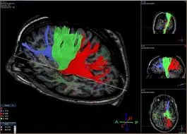
Die Diffusionstensorbildgebung bietet eine umfassende Sicht auf neuronale Wege und liefert detaillierte Bilder von weißen Substanzbahnen und der Gehirnvernetzung. Dieser Abschnitt umreißt, was DTI ist und welche Vorteile es für die Diagnose verschiedener Erkrankungen hat.
Was zeigt die Diffusionstensorbildgebung?
- Weiße Substanzbahnen: Überlegene Bildgebung der weißen Substanzwege des Gehirns.
- Gehirnvernetzung: Klare Visualisierung neuronaler Verbindungen, nützlich bei der Diagnose von Vernetzungsproblemen.
- Mikrostrukturelle Veränderungen: Erkennt Veränderungen innerhalb der weißen Substanz, die in herkömmlichen MRTs nicht sichtbar sind.
- Krankheitserkennung: Effektiver bei der Identifizierung von Erkrankungen wie Multipler Sklerose und traumatischen Hirnverletzungen.
Wann werden Sie eine erhalten?
- Persistierende neurologische Symptome: Besonders wenn sie durch herkömmliche MRT nicht ausreichend erklärt werden können.
- Neurodegenerative Erkrankungen: Wie Alzheimer oder Multiple Sklerose.
- Präoperative Planung: Bietet eine detaillierte Karte für die chirurgische Planung bei Gehirnoperationen.
- Forschung: Ideal zur Untersuchung der Gehirnvernetzung und mikrostruktureller Veränderungen in Forschungseinrichtungen.
Techniken zur Interpretation der Diffusionstensorbildgebung
Die Interpretation von DTI erfordert ein tieferes Verständnis fortgeschrittener Bildgebungstechniken. Hier sind drei Techniken für Fachleute auf unterschiedlichen Erfahrungsstufen.
1. Nutzung von X-ray Interpreter
X-ray Interpreter erweitert jetzt seine KI-gesteuerte Analyse auf DTI-Bilder. Der Prozess sichert Präzision:
- Registrierung: Registrieren Sie sich auf X-ray Interpreter, um auf die KI-Analyse für DTI zuzugreifen.
- Hochladen von DTIs: Laden Sie Ihre DTI-Bilder hoch.
- Überprüfung der Interpretation: Erhalten Sie die KI-generierte Interpretation und laden Sie Ihren Bericht herunter.
- Professionelle Beratung: Es ist immer ratsam, sich mit medizinischen Fachleuten zu beraten, um ein umfassendes Verständnis zu erhalten.
Bitte sehen Sie sich unseren Leitfaden zum Einstieg an.
2. Verwendung von ChatGPT Plus
ChatGPT Plus unterstützt jetzt die Analyse von DTI-Bildern mit seinem neuesten GPT-4V-Modell, das detaillierte und interaktive Einblicke bietet:
- Abonnement: Abonnieren Sie ChatGPT Plus für erweiterte Bildanalysefunktionen.
- Hochladen von DTIs: Kommunizieren Sie mit GPT-4V auf OpenAI, um Ihre DTI-Bilder hochzuladen.
- Anfordern von Analysen: Arbeiten Sie mit dem Modell für eine gründliche Analyse.
- Überprüfung und Bestätigung: Bewerten und verfeinern Sie die Analyse nach Bedarf.
- Professionelle Validierung: Eine Validierung durch medizinische Experten wird empfohlen.
Weitere Informationen finden Sie in unserem Beitrag über die Verwendung von ChatGPT Plus zur MRI-Interpretation.
Alternativ können Sie, während mehrere andere KI-Modelle mit visuellen Fähigkeiten auftauchen, auch andere Modelle ausprobieren, wie Grok von xAI, Claude von Anthropic, Gemini von Google Deepmind.
3. Meistere die DTI-Interpretation selbst
Für Gesundheitsdienstleister, die ihre Fähigkeiten im DTI-Lesen verbessern möchten, ist Selbstlernen von unschätzbarem Wert:
- Bildung: Verfolgen Sie eine fortgeschrittene Ausbildung in DTI-Interpretation.
- Übung: Regelmäßige Praxis unter fachkundiger Anleitung.
- Ressourcen: Nutzen Sie Bücher zur fortgeschrittenen Bildgebung und Online-Kurse.
- Feedback: Suchen Sie nach Feedback, um Fähigkeiten zu verfeinern.
- Lebenslanges Lernen: Beteiligen Sie sich an fortlaufender Bildung, um mit den Bildgebungstechniken aktuell zu bleiben.
Empfohlene Ressourcen für das Selbstlernen:
-
Diffusionstensorbildgebung und Faserverfolgung - Radiopaedia: Bietet einen umfassenden Überblick über DTI und Faserverfolgung, einschließlich ihrer Anwendungen, Einschränkungen und der Wissenschaft hinter der Technik.
-
Diffusionstensorbildgebung - YouTube: Ein detailliertes Video, das die Prinzipien von DTI, seine klinischen Anwendungen und die Visualisierung neuronaler Wege im Gehirn erklärt.
-
Diffusionstensorbildgebung - ScienceDirect: Bietet tiefgehende Artikel und Forschungsarbeiten zu DTI, die dessen Methodik, klinische Anwendungen und aktuelle Fortschritte im Feld abdecken.
Vergleichende Analyse
Die Wahl der richtigen Technik zur Interpretation von DTI ist entscheidend für eine genaue Diagnose. Dieser Abschnitt vergleicht die drei Methoden:
| Kriterien | X-ray Interpreter | ChatGPT Plus | Selbstlesung |
|---|---|---|---|
| Genauigkeit | Hoch (KI-basiert)1 | Hoch (KI-basiert)1 | Variiert (Fähigkeitsabhängig) |
| Benutzerfreundlichkeit | Einfach | Mäßig | Herausfordernd |
| Kosten | Ab 2,50 $ pro Bild | 20 $ pro Monat | Kostenlos (außer Bildungskosten) |
| Zeiteffizienz | Schnell | Mäßig bis Schnell | Langsam bis Mäßig |
| Lernkurve | Niedrig | Niedrig bis Mäßig | Hoch |
| Zusätzliche Ressourcen | Bereitgestellt | Teilweise bereitgestellt (über OpenAI) | Selbstbeschafft |
Jede Methode hat ihre Stärken und Schwächen, wobei KI-Optionen schnelle und präzise Interpretationen bieten, während das Selbstlesen eine tiefgehende Lernmöglichkeit für medizinische Fachkräfte fördert.
Fazit
Die DTI-Interpretation ist entscheidend für die Diagnose komplexer neurologischer Erkrankungen. Dieser Leitfaden präsentiert drei Techniken, die auf unterschiedliche professionelle Bedürfnisse und Kompetenzstufen abgestimmt sind. KI-Methoden bieten schnelle, präzise Interpretationen, während das Selbstlernen auf diejenigen abzielt, die an tiefgehenden Kenntnissen interessiert sind.
Bei der Auswahl einer Technik sollten Sie Ihr Erfahrungsniveau, den Bedarf an zeitgerechter Interpretation und die verfügbaren Ressourcen berücksichtigen. Ethische und rechtliche Standards müssen immer eingehalten werden, um die Sicherheit und Vertraulichkeit der Patienten zu gewährleisten.
Ressourcen und Weiterführendes Lernen
Für eine weitere Erkundung und ein besseres Verständnis der DTI-Interpretation ziehen Sie die folgenden Ressourcen in Betracht:
-
Diffusionstensorbildgebung - Medscape: Bietet einen gründlichen Überblick über DTI, einschließlich seiner klinischen Anwendungen, Techniken und Einschränkungen.
-
Magnetresonanztomographie des Gehirns - Wiley Online Library: Eine akademische Arbeit, die verschiedene MRT-Techniken, einschließlich DTI, und deren Anwendungen in der medizinischen Bildgebung diskutiert.
-
Funktionelle MRT und Diffusionstensorbildgebung - Mayfield Clinic: Eine informative Ressource zur Verwendung von fMRT und DTI zur Visualisierung von Gehirnaktivität und weißen Substanzbahnen.