كيفية تفسير الموجات فوق الصوتية الوعائية: 3 طرق بسيطة
تعتبر الموجات فوق الصوتية الوعائية تقنية تصوير غير جراحية تستخدم الموجات الصوتية لتقييم تدفق الدم في شرايينك وأوردةك. يشرح هذا الدليل ما يمكن أن تكشفه الموجات فوق الصوتية الوعائية، ولماذا تُستخدم، وكيفية تفسير نتائجها بشكل فعال.
ما هي الموجات فوق الصوتية الوعائية؟
تستخدم الموجات فوق الصوتية الوعائية موجات صوتية عالية التردد لإنشاء صور للأوعية الدموية. على عكس الأشعة السينية، لا تستخدم الإشعاع، مما يجعلها خيارًا آمنًا وغير مؤلم. دعونا نستكشف ما تتضمنه هذه التقنية وما تظهره.
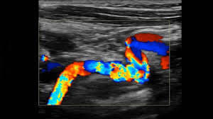
ماذا يمكن أن تظهر الموجات فوق الصوتية الوعائية؟
- تحليل تدفق الدم: يقيس سرعة واتجاه تدفق الدم.
- هيكل الأوعية: تصور جدران وحجم الشرايين والأوردة.
- كشف الانسداد: تحدد الأوعية الضيقة أو المسدودة بسبب الخثرات أو اللويحات.
- كشف الأنيوريسم: تساعد في العثور على انتفاخات غير طبيعية في الأوعية الدموية.
- التصوير في الوقت الحقيقي: يوفر مشاهد ديناميكية لأنماط تدفق الدم.
لماذا قد تحتاج إلى الموجات فوق الصوتية الوعائية؟
- ألم أو تورم في الساق: للتحقق من وجود تجلط الأوردة العميقة (DVT).
- تقييم خطر السكتة الدماغية: لتقييم الشرايين السباتية للضيق.
- مرض الشرايين المحيطية (PAD): لتقييم تدفق الدم في الأطراف.
- اشتباك الأنيوريسم: لتحديد الانتفاخات المحتملة في الأوعية.
- التقييم قبل الجراحة: لتقييم صحة الأوعية قبل الإجراءات الجراحية.
كيف تُجرى الموجات فوق الصوتية الوعائية؟
الموجات فوق الصوتية الوعائية إجراء آمن وغير جراحي. إليك ما يمكن أن تتوقعه:
- التحضير: سيُطلب منك الاستلقاء، عادةً على طاولة الفحص. ستتعرض المنطقة التي يتم فحصها (مثل الساق، الذراع، البطن).
- تطبيق الجل: سيتم تطبيق جل شفاف قائم على الماء على بشرتك فوق منطقة الأوعية الدموية التي سيتم فحصها. يساعد هذا الجل في تحسين الاتصال بين المحول والجلد.
- تحريك المحول: سيقوم فني الموجات فوق الصوتية بتحريك المحول بلطف على بشرتك. ينبعث المحول موجات صوتية لإنشاء صور للأوعية الدموية.
- اكتساب الصور: تلتقط آلة الموجات فوق الصوتية صورًا في الوقت الحقيقي للأوعية الدموية وتدفق الدم. يتيح ذلك للفني تقييم حجم وشكل الأوعية واتجاه وسرعة تدفق الدم.
- الموجات فوق الصوتية دوبلر: غالبًا ما تُستخدم تقنية الموجات فوق الصوتية دوبلر لتقييم تدفق الدم. يمكن أن تساعد في الكشف عن الانسداد أو الضيق أو أنماط التدفق غير الطبيعية.
- المدة: يمكن أن تختلف مدة الفحص حسب المنطقة التي يتم فحصها، لكن عادةً ما تستغرق حوالي 30 إلى 60 دقيقة.
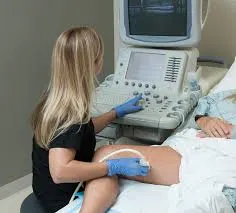
الإجراء غير مؤلم ويمكنك عادةً العودة إلى أنشطتك الطبيعية على الفور.
كيفية تفسير نتائج الموجات فوق الصوتية الوعائية
فهم نتائج الموجات فوق الصوتية الوعائية أمر حاسم لإدارة صحتك الوعائية. إليك ثلاث طرق مختلفة للمساعدة في تفسير النتائج:
1. استخدام X-ray Interpreter
يقدم X-ray Interpreter الآن تحليلًا مدفوعًا بالذكاء الاصطناعي لصور الموجات فوق الصوتية الوعائية. إليك كيفية استخدامه:
- التسجيل: قم بالتسجيل على X-ray Interpreter لاستخدام الذكاء الاصطناعي لدينا للتحليل.
- تحميل الموجات فوق الصوتية: قم بتحميل صور الموجات فوق الصوتية الوعائية الخاصة بك.
- مراجعة التفسير: استقبل التفسير المستند إلى الذكاء الاصطناعي، بما في ذلك تقرير.
- استشارة: استشر دائمًا طبيبك للحصول على تشخيص شامل.
تحقق من دليل بدء الاستخدام الخاص بنا لمزيد من التفاصيل.
2. استخدام ChatGPT Plus
يمكن أن تساعد ChatGPT Plus، باستخدام نموذج GPT-4V المتقدم، في تحليل صور الموجات فوق الصوتية الوعائية:
- الاشتراك: اشترك في ChatGPT Plus للحصول على تحليلات متقدمة.
- تحميل الموجات فوق الصوتية: قم بتحميل صور الموجات فوق الصوتية الوعائية الخاصة بك على منصة OpenAI.
- طلب التحليل: اطلب من الذكاء الاصطناعي تفسير صور الموجات فوق الصوتية الخاصة بك وتقديم تقرير.
- مراجعة وتأكيد: قم بمراجعة النتائج وتأكيد دقتها مع محترف الرعاية الصحية.
اكتشف المزيد في مدونتنا حول استخدام ChatGPT Plus لتفسير الصور الطبية.
بدلاً من ذلك، مع ظهور العديد من نماذج الذكاء الاصطناعي الأخرى ذات القدرات البصرية، يمكنك أيضًا تجربة نماذج أخرى، مثل Grok من xAI، Claude من Anthropic، Gemini من Google Deepmind.
3. فهم الأساسيات بنفسك
بينما لا تعتبر بديلاً عن المتخصصين في الرعاية الصحية، يمكن أن تساعدك معرفة بعض الأساسيات في فهم النتائج بشكل أفضل وتحضير الأسئلة لطبيبك.
- تعلم تشريح الأوعية: تعرف على تشريح الأوعية الدموية الأساسية.
- قراءة أدلة بسيطة: يمكن أن تساعدك العديد من المصادر على الإنترنت في فهم النتائج الشائعة في الموجات فوق الصوتية الوعائية.
- طرح الأسئلة: قم بتدوين أي مصطلحات غير مألوفة واسأل مقدم الرعاية الصحية لديك خلال المتابعة.
- السعي للحصول على إرشادات الخبراء: تحقق دائمًا من فهمك مع ممارس طبي محترف.
مقارنة الأساليب المختلفة
دعونا نقارن الطرق المختلفة لتفسير الموجات فوق الصوتية الوعائية:
| المعايير | X-ray Interpreter | ChatGPT Plus | القراءة الذاتية |
|---|---|---|---|
| الدقة | عالية (مبنية على الذكاء الاصطناعي)1 | عالية (مبنية على الذكاء الاصطناعي)1 | تختلف (تعتمد على المهارات) |
| سهولة الاستخدام | سهلة | معتدلة | تحدي |
| التكلفة | بدءًا من 2.50 دولار لكل صورة | 20 دولار في الشهر | مجانية (باستثناء التكاليف التعليمية) |
| كفاءة الوقت | سريعة | معتدلة إلى سريعة | بطيئة إلى معتدلة |
| منحنى التعلم | منخفض | منخفض إلى معتدل | مرتفع |
| موارد إضافية | مقدمة | جزئياً مقدمة (من خلال OpenAI) | مستمدة ذاتياً |
كل طريقة لها فوائدها وقيودها. تقدم الخيارات المدفوعة بالذكاء الاصطناعي سرعة ودقة، بينما تساعد المعرفة الأساسية في تحسين التواصل بين المريض والطبيب.
الخاتمة
تعتبر الموجات فوق الصوتية الوعائية ضرورية لتشخيص العديد من الحالات الوعائية. قدّم لك هذا الدليل تقنية الموجات فوق الصوتية الوعائية، والصور التي تنتجها، وكيف يمكنك فهم نتائجك بشكل أفضل من خلال أدوات الذكاء الاصطناعي والبحث الذاتي.
عند اختيار الطريقة، ضع في اعتبارك احتياجاتك المحددة، ومستوى الفهم المرغوب فيه، والموارد المتاحة. دائمًا ما يجب إعطاء الأولوية للخصوصية وطلب التحقق الطبي المحترف.
مقالات ذات صلة
- كيفية تفسير تخطيطات القلب بالأمواج فوق الصوتية
- كيفية تفسير تصوير الأوعية المقطعية
- كيفية تفسير التصوير بالأشعة المغناطيسية للأوعية