如何解读盆腔超声:三种基本方法
December 13, 2024
盆腔超声是一种非侵入性成像技术,使用声波可视化盆腔器官。 本指南解释了盆腔超声可以揭示的内容、其用途以及如何理解其发现。
盆腔超声是什么?
盆腔超声使用声波实时创建盆腔内器官和组织的图像。 安全且无痛,不使用辐射,使其成为宝贵的诊断工具。
盆腔超声可以显示什么?
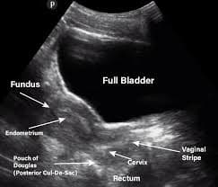
- 女性生殖器官: 清晰的子宫、卵巢和宫颈图像。
- 膀胱: 膀胱及其周围结构的可视化。
- 液体检测: 识别囊肿或游离液体等液体积聚。
- 异常识别: 检测肿块、肿瘤、肌瘤和卵巢囊肿。
- 实时成像: 让医生评估器官功能和血流。
你为什么可能需要盆腔超声?
- 盆腔疼痛: 调查不明盆腔疼痛的原因。
- 月经不规律: 评估异常出血或周期。
- 怀疑有肌瘤或囊肿: 检查肿块或囊肿。
- 怀孕监测: 监测胎儿的发育。
- 不孕评估: 作为生育问题检査的一部分。
盆腔超声如何进行?
盆腔超声是一种简单的程序,通常需要15到30分钟。以下是你可以期待的内容:
准备工作
- 衣物: 可能会要求你更换为医用服装。穿着舒适、宽松的衣物以便于操作。
- 充满膀胱(通常): 对于某些类型的盆腔超声,特别是经腹超声,充满膀胱是必要的,以提供更好的盆腔器官视图。医生可能要求你在扫描前饮水。如果需要,技术员会给出指示。
程序
- 涂抹凝胶: 在下腹部或盆腔区域涂上一层透明的水基凝胶。这有助于换能器与皮肤良好接触。
- 换能器移动: 超声技师将一种称为换能器的手持设备在涂抹凝胶的区域上移动。换能器发出声波,这些声波从器官和组织反弹,形成图像。
- 图像采集: 超声技师在显示器上观察图像。可能会拍摄快照或短视频以进行详细分析。
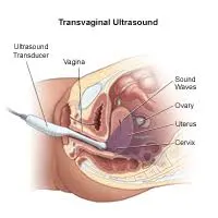
- 经阴道超声(如有需要): 在某些情况下,可能需要进行经阴道超声,以更清晰地查看子宫和卵巢。一个小的润滑换能器将轻柔地插入阴道。这不是疼痛的,类似于刷宫颈涂片。
超声后
- 凝胶去除: 将凝胶擦去后,你可以穿衣服。
- 结果: 图像由放射科医生或你的医生解读。通常你会在后续预约时收到结果。
该程序通常无痛且非常安全。如果你有疑问或担忧,请随时向超声技师或你的医疗提供者询问。
如何解读盆腔超声结果
理解你的盆腔超声结果可以帮助你更好地管理你的健康。以下是一些更好地解读结果的方法。
1. 利用X-ray Interpreter
X-ray Interpreter提供超声图像的AI驱动分析:
- 注册: 在X-ray Interpreter上注册以获得超声分析。
- 上传超声: 上传你的盆腔超声图像。
- 审查解读: 收到AI生成的解读报告。
- 咨询: 始终与医生咨询以获得诊断。
请查看我们的入门指南以获取更多细节。
2. 使用ChatGPT Plus
ChatGPT Plus(GPT-4V)可以帮助分析超声图像:
- 订阅: 订阅ChatGPT Plus以获得高级分析。
- 上传超声: 在OpenAI平台上上传图像。
- 请求分析: 请AI解读你的超声图像。
- 审核和验证: 与医疗专业人员一起审核和确认准确性。
在我们的博客中了解更多关于如何使用ChatGPT Plus进行医学图像解读的信息。
此外,随着其他具有视觉能力的AI模型的出现,你还可以尝试其他模型,例如 由xAI提供的Grok, 由Anthropic提供的Claude, 或由Google Deepmind提供的Gemini。
3. 自己理解基础知识
理解基础知识可以帮助你对医学有更深入的了解,但这并不能替代医学专业知识。
- 学习解剖学: 熟悉盆腔器官的解剖结构。
- 阅读简单指南: 在线资源可以帮助你理解常见的超声发现。
- 提问: 向医疗服务提供者询问不熟悉的术语。
- 寻求专家指导: 始终与医疗专业人员验证你的理解。
比较不同的方法
让我们比较一下盆腔超声的不同解读方法:
| 标准 | X-ray Interpreter | ChatGPT Plus | 自我阅读 |
|---|---|---|---|
| 准确性 | 高(基于AI)1 | 高(基于AI)1 | 变化(依赖于技能) |
| 易用性 | 容易 | 中等 | 挑战性 |
| 成本 | 每张图像起价2.50美元 | 每月20美元 | 免费(不包括教育费用) |
| 时间效率 | 快 | 中等到快 | 慢到中等 |
| 学习曲线 | 低 | 低到中等 | 高 |
| 额外资源 | 提供 | 部分提供(通过OpenAI) | 自行搜集 |
每种方法都有其优点。AI选项提供速度和精确度,而基础理解可以改善与医生的沟通。
结论
盆腔超声对诊断各种疾病至关重要。 本指南向你介绍了如何解读和理解你的结果。
在选择方法时,请考虑你的需求、理解水平和可用资源。始终寻求专家的验证,并遵守隐私标准。