Comment interpréter les échographies scrotales : 3 Méthodes Essentielles
Une échographie scrotale est une technique d'imagerie clé utilisant des ondes sonores pour examiner les testicules et les tissus environnants. Ce guide explique ce que cette échographie peut révéler, pourquoi elle est nécessaire et comment interpréter ses résultats.
Qu'est-ce qu'une Échographie Scrotale ?
Une échographie scrotale est un test d'imagerie non invasif qui fournit des images en temps réel des testicules, de l'épididyme et des structures environnantes. Utilisant des ondes sonores, c'est un moyen sûr et indolore d'évaluer les organes reproducteurs masculins. Explorons ce qu'elle montre et le processus.
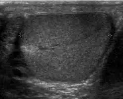
Que Peut Montrer une Échographie Scrotale ?
- Imagerie Testiculaire Détailée : Visualisation claire des testicules, de l'épididyme et des cordons spermatiques.
- Détection de Fluide : Détecte les collections de fluides comme les hydrocèles ou les varicocèles.
- Identification d'Anomalies : Aide à identifier les masses, tumeurs, kystes et signes d'inflammation ou d'infection.
- Évaluation du Flux Sanguin : Évalue le flux sanguin vers les testicules, crucial pour le diagnostic de la torsion.
- Imagerie en Temps Réel : Permet aux médecins d'observer les structures en temps réel.
Pourquoi Pourriez-Vous Avoir Besoin d'une Échographie Scrotale ?
- Douleur ou Gonflement Testiculaire : Pour identifier la cause de l'inconfort ou de l'enflure.
- Masse ou Nodule Palpable : Pour évaluer toute anomalie ressentie lors des auto-examens ou par un médecin.
- Préoccupations en Matière d'Infertilité : Pour évaluer la structure testiculaire et identifier d'éventuels problèmes.
- Traumatisme du Scrotum : Pour évaluer les dommages après une blessure.
- Torsion Suspectée : Pour écarter ou confirmer la torsion testiculaire, une urgence médicale.
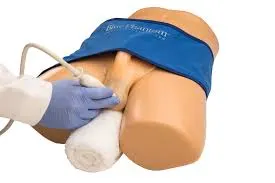
Comment est Réalisée une Échographie Scrotale ?
Une échographie scrotale est une procédure rapide, indolore et non invasive. Voici ce à quoi vous pouvez vous attendre :
-
Préparation : On vous demandera de retirer tout vêtement du bas et vous sera fourni une blouse ou un drap. Vous vous allongerez ensuite sur le dos sur une table d'examen.
-
Application du Gel : Un gel clair à base d'eau sera appliqué sur la peau de votre scrotum. Ce gel aide le transducteur (le dispositif manuel qui émet des ondes sonores) à bien entrer en contact avec votre peau et permet une meilleure transmission des ondes sonores.
-
Mouvement du Transducteur : Le sonographe (le professionnel formé à réaliser l'échographie) déplacera doucement le transducteur sur la zone recouverte de gel de votre scrotum. Il appliquera une légère pression au besoin pour obtenir les meilleures images des testicules, de l'épididyme et des structures environnantes.
-
Acquisition d'Images : Au fur et à mesure que le transducteur est déplacé, des images en temps réel sont affichées sur un moniteur. Le sonographe peut prendre des images fixes ou des clips vidéo de zones particulières. On pourrait vous demander de rester immobile ou de changer votre position légèrement pour obtenir les meilleures vues.
-
Durée de la Procédure : La procédure entière dure généralement environ 15 à 30 minutes.
-
Post-Procedé : Après la procédure, le gel sera essuyé, et vous pourrez vous habiller et retourner à vos activités. Les images sont ensuite généralement examinées par un radiologue, qui fournira un rapport à votre médecin.
Comment Interpréter les Résultats de l'Échographie Scrotale
Comprendre vos résultats d'échographie est crucial pour gérer votre santé. Voici comment vous pouvez obtenir de l'aide pour les interpréter.
1. Utilisation de X-ray Interpreter
X-ray Interpreter fournit maintenant une analyse basée sur l'IA pour les images d'échographie, y compris les échographies scrotales. Voici comment l'utiliser :
- Inscription : Inscrivez-vous sur X-ray Interpreter pour utiliser l'IA pour l'analyse des échographies.
- Téléchargement d'Échographies : Téléversez vos images d'échographie scrotale.
- Examen de l'Interprétation : Obtenez une analyse générée par l'IA et un rapport détaillé.
- Consultation : Consultez toujours votre médecin pour un diagnostic complet.
Vérifiez notre guide de démarrage pour plus de détails.
2. Utilisation de ChatGPT Plus
ChatGPT Plus, utilisant GPT-4V, peut aider à analyser les images d'échographie :
- Abonnement : Abonnez-vous à ChatGPT Plus pour une analyse avancée.
- Téléchargement d'Échographies : Téléversez vos images sur la plateforme OpenAI.
- Demande d'Analyse : Demandez à l'IA d'interpréter votre échographie et de fournir un rapport.
- Examen et Validation : Examinez les résultats avec un professionnel de la santé.
Découvrez plus dans notre blog sur l'utilisation de ChatGPT Plus pour l'interprétation d'images médicales.
Alternativement, alors que plusieurs autres modèles d'IA avec des capacités visuelles émergent, vous pouvez également essayer d'autres modèles, tels que Grok de xAI, Claude de Anthropic, Gemini de Google Deepmind.
3. Comprendre les Bases Vous-Même
Bien que cela ne remplace pas les conseils médicaux, comprendre des concepts de base peut vous aider à mieux comprendre les résultats.
- Apprenez l'Anatomie : Familiarisez-vous avec l'anatomie des testicules, de l'épididyme et des structures environnantes.
- Lisez des Guides : De nombreuses ressources en ligne expliquent des résultats d'échographie communs.
- Posez des Questions : Notez les termes inconnus et clarifiez avec votre fournisseur de soins.
- Cherchez des Conseils d'Expert : Validez toujours votre compréhension avec un professionnel médical.
Comparaison des Différentes Approches
Comparons les méthodes pour interpréter les échographies scrotales :
| Critères | X-ray Interpreter | ChatGPT Plus | Auto-Lecture |
|---|---|---|---|
| Précision | Élevée (basée sur l'IA)1 | Élevée (basée sur l'IA)1 | Varie (dépend des compétences) |
| Facilité d'Utilisation | Facile | Modérée | Difficile |
| Coût | À partir de 2,50 $ par image | 20 $ par mois | Gratuit (hors frais éducatifs) |
| Efficacité Temporelle | Rapide | Modérée à Rapide | Lent à Modéré |
| Courbe d'Apprentissage | Faible | Faible à Modérée | Élevée |
| Ressources Supplémentaires | Fournies | Partiellement Fournies (via OpenAI) | Auto-sourcées |
Les méthodes basées sur l'IA offrent rapidité et précision, tandis que des connaissances de base favorisent une meilleure communication avec les médecins.
Conclusion
Les échographies scrotales sont essentielles pour diagnostiquer les problèmes reproducteurs masculins. Ce guide a expliqué leur utilisation, les images qu'elles produisent et les méthodes pour comprendre les résultats en utilisant des outils d'IA et l'auto-éducation.
Lors du choix d'une méthode, tenez compte de vos besoins spécifiques et de vos ressources. Priorisez toujours la confidentialité et cherchez une validation professionnelle.